. 2012 Feb 7;109(6):2132-7.
doi: 10.1073/pnas.1118847109. Epub 2012 Jan 23.

Gene therapy rescues photoreceptor blindness in dogs and paves the way for treating human X-linked retinitis pigmentosa

Affiliations

Gene therapy rescues photoreceptor blindness in dogs and paves the way for treating human X-linked retinitis pigmentosa

William A Beltran et al. Proc Natl Acad Sci U S A. .

Abstract

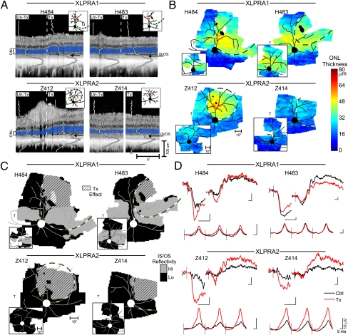
Hereditary retinal blindness is caused by mutations in genes expressed in photoreceptors or retinal pigment epithelium. Gene therapy in mouse and dog models of a primary retinal pigment epithelium disease has already been translated to human clinical trials with encouraging results. Treatment for common primary photoreceptor blindness, however, has not yet moved from proof of concept to the clinic. We evaluated gene augmentation therapy in two blinding canine photoreceptor diseases that model the common X-linked form of retinitis pigmentosa caused by mutations in the retinitis pigmentosa GTPase regulator (RPGR) gene, which encodes a photoreceptor ciliary protein, and provide evidence that the therapy is effective. After subretinal injections of adeno-associated virus-2/5-vectored human RPGR with human IRBP or GRK1 promoters, in vivo imaging showed preserved photoreceptor nuclei and inner/outer segments that were limited to treated areas. Both rod and cone photoreceptor function were greater in treated (three of four) than in control eyes. Histopathology indicated normal photoreceptor structure and reversal of opsin mislocalization in treated areas expressing human RPGR protein in rods and cones. Postreceptoral remodeling was also corrected: there was reversal of bipolar cell dendrite retraction evident with bipolar cell markers and preservation of outer plexiform layer thickness. Efficacy of gene therapy in these large animal models of X-linked retinitis pigmentosa provides a path for translation to human treatment.


Conflict of interest statement

Conflict of interest statement: The authors declare a conflict of interest. W.W.H. and the University of Florida have a financial interest in the use of adeno-associated virus therapies and own equity in a company (AGTC Inc.) that might, in the future, commercialize some aspects of this work. The remaining authors declare no conflict of interest.

Figures

Fig. 1.
Fig. 1.
Retinal disease phenotypes caused by RPGRORF15 mutations in human patients and in dogs. (A) Different patterns of photoreceptor topography in two XLRP patients with RPGR mutations (P1: c.ORF15+483_484delGA, p.E746fs; P2: c.ORF15+ 652_653delAG, p.E802fs). ONL thickness topography is mapped to a pseudocolor scale. (Inset) Representative normal subject. Location of fovea and optic nerve (ON) are shown. (B) Different patterns of photoreceptor topography in the canine models of RPGRORF15; mapping as performed with the human data. (Inset) Map of a representative WT dog with location of ON labeled. (C) ONL thickness profile along the vertical meridian (Inset) comparing XLPRA1 and XLPRA2 of different ages (thin traces) versus normal results (gray band). Mean (±SD) results are from groups of younger (7–28 wk) and older (36–76 wk) dogs. The thicker red line represents the data from the oldest dogs examined (>144 wk old). Brackets mark the location of the high photoreceptor density corresponding to the canine visual streak. (D) Rod and cone retinal function by ERGs in XLPRA1 (young: 7–23 wk; old: 56–80 wk) and XLPRA2 (young: 8–22 wk; old: 38–144 wk) dogs shown as the logarithm of amplitude loss from the mean WT value (rod: 2.39 and 2.38 log10 μV and cone: 1.50 and 1.72 log10 μV for younger and older, respectively). Each symbol represents an eye. Horizontal dashed lines represent the WT limits (±2 SD).
Fig. 2.
Fig. 2.
In vivo evidence of gene augmentation therapy success in XLPRA dogs. (A) Cross-sectional OCT retinal scans crossing the treatment bleb boundary (dashed line in H484, H483, and Z412) or comparing inside and outside the bleb region (white space in Z414) in treated eyes of XLPRA1 (H484, H483) and XLPRA2 (Z412, Z414) dogs. ONL is highlighted in blue for visibility. Overlaid are the longitudinal reflectivity profiles defining the backscattered light intensity from different retinal layers. Arrows point to the backscatter peak originating from the IS/OS region. (Insets) Red line represents the location of the scans. (B) Topography of ONL thickness in treated eyes shown on a pseudocolor scale with superimposed retinal blood vessels and optic nerve. White represents no data; irregularly shaped black foci indicate retinotomy sites. Bleb boundaries are outlined with green-and-white dashed lines. Small inset figures are BSS-treated control fellow eyes. (C) Topography of average backscatter intensity originating from the photoreceptor IS/OS region in treated eyes with superimposed retinal blood vessels and optic nerve. The same threshold is used in all eyes to distinguish regions of high (gray) and low (black) IS/OS backscatter. Diagonal-pattern regions delineate the treatment effect by comparison of the two eyes. All eyes are shown as equivalent right eyes for comparability. T, temporal retina. (B and C) (Insets) BSS-treated contralateral eyes. (D) ERGs in treated (red traces) and BSS-injected control fellow eyes (black traces). For each panel in D, the upper-left waveforms are the leading edges of the photoresponses driven by rod photoreceptor activation, and the upper-right waveforms are the b-waves dominated by rod bipolar cells, both recorded under dark-adapted conditions. Lower waveforms are 29-Hz flicker responses dominated by cone function recorded under light-adapted conditions. Black vertical lines show the timing of flash onset. Calibrations are 5 ms (abscissa) and 10 μV (ordinate); note the ∼3× larger waveforms of H483.
Fig. 3.
Fig. 3.
Gene augmentation therapy rescues photoreceptors in the XLPRA1 dog H484 treated with AAV2/5-hIRBP-hRPGR at 28 wk of age and terminated at 77 wk. The schematic drawing illustrates the treatment area (dashed green lines) and the location of the region (red line) illustrated in the section. (1) Representative H&E-stained cryosection at the nontreated/treated junction (vertical dashed line). Boxed areas are illustrated at higher magnification below (2–5). Photoreceptor density is decreased in nontreated region and both ONL (white arrowheads) and OPL are narrowed; rod and cone IS are short, and OS sparse. In treated regions, the number of photoreceptors is increased and their structure is normal (4 and 5), resulting in thicker ONL and preserved OPL. (68) Expression of hRPGRORF15 in treated areas decreases in the transition zone and is absent elsewhere. Protein is present in rod and cone inner segments and synaptic regions and, to a lesser extent, in the perinuclear cytoplasm where expression is most intense. (9,10,12, and 13) Rod (RHO) and red/green cone (R/G ops) opsins are mislocalized in untreated regions with label in the IS, ONL, and synaptic terminals. Treated areas show normal localization to the OS. (11 and 14) Preservation of normal cone structure in treated areas is clearly shown with cone arrestin (Cone Arr) labeling. GCL, ganglion cell layer; INL, inner nuclear layer; IS, inner segments; ONL, outer nuclear layer; OPL, outer plexiform layer; OS, outer segments; RPE, retinal pigment epithelium.
Fig. 4.
Fig. 4.
Successful gene therapy rescues retinal remodeling in the XLPRA2 dog Z412 treated with AAV2/5-hIRPB-hRPGR at 5 wk of age and terminated at 38 wk. Immunolabeling with CtBP2/RIBEYE shows a reduced number of photoreceptor synaptic ribbons in the untreated areas (1). In treated areas, the density of synaptic ribbons is normal, thus contributing to the preservation of the OPL thickness (2). Coimmunolabeling of rod bipolar (PKCα) and ON bipolar cells (Goα) shows retraction of dendrites in untreated areas (3), whereas dendritic arborization is preserved in treated regions (4). (5 and 6) Coimmunolabeling of the inner retina with antibodies to neurofilament 200 kDa (NF200) and calbindin (Calb) is normal in both untreated and treated regions, but punctate NF200 staining is seen in the ONL in untreated areas. (7 and 8) GFAP immunolabeling of Müller cell radial extensions is found only in untreated areas, whereas no reactive Müller cells are seen in the treated regions.

References

    1. Wright AF, Chakarova CF, Abd El-Aziz MM, Bhattacharya SS. Photoreceptor degeneration: Genetic and mechanistic dissection of a complex trait. Nat Rev Genet. 2010;11:273–284. - PubMed
    1. Bramall AN, Wright AF, Jacobson SG, McInnes RR. The genomic, biochemical, and cellular responses of the retina in inherited photoreceptor degenerations and prospects for the treatment of these disorders. Annu Rev Neurosci. 2010;33:441–472. - PubMed
    1. Acland GM, et al. Gene therapy restores vision in a canine model of childhood blindness. Nat Genet. 2001;28:92–95. - PubMed
    1. Cideciyan AV. Leber congenital amaurosis due to RPE65 mutations and its treatment with gene therapy. Prog Retin Eye Res. 2010;29:398–427. - PMC - PubMed
    1. Bird AC. X-linked retinitis pigmentosa. Br J Ophthalmol. 1975;59:177–199. - PMC - PubMed

Publication types

MeSH terms