. 2012 Feb 7;109(6):2168-73.
doi: 10.1073/pnas.1119229109. Epub 2012 Jan 19.

Ecotopic viral integration site 1 (EVI1) regulates multiple cellular processes important for cancer and is a synergistic partner for FOS protein in invasive tumors

Affiliations

Ecotopic viral integration site 1 (EVI1) regulates multiple cellular processes important for cancer and is a synergistic partner for FOS protein in invasive tumors

Emilie A Bard-Chapeau et al. Proc Natl Acad Sci U S A. .

Abstract

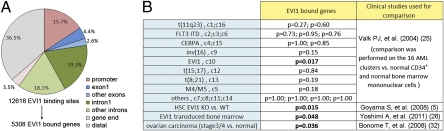
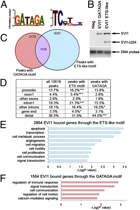
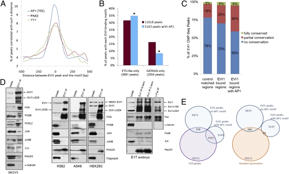
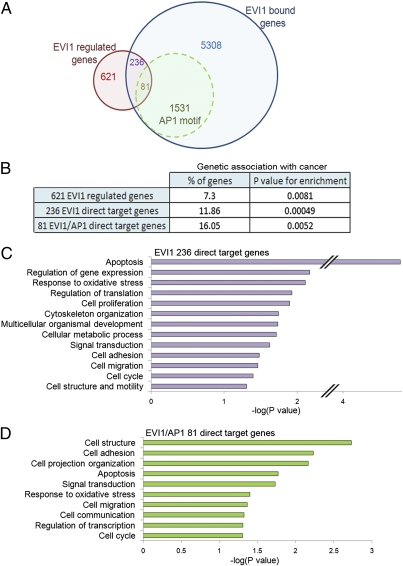
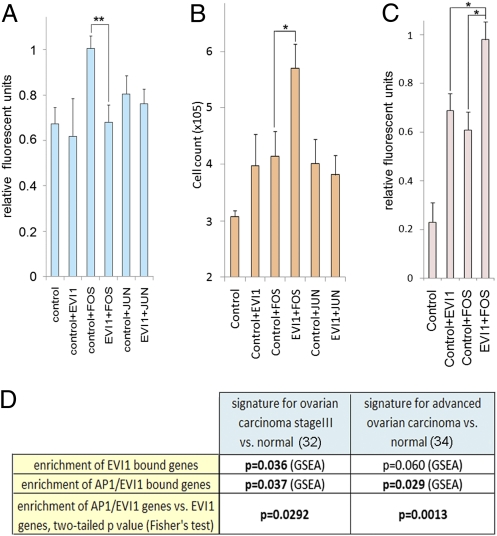
Ecotropic viral integration site 1 (EVI1) is an oncogenic dual domain zinc finger transcription factor that plays an essential role in the regulation of hematopoietic stem cell renewal, and its overexpression in myeloid leukemia and epithelial cancers is associated with poor patient survival. Despite the discovery of EVI1 in 1988 and its emerging role as a dominant oncogene in various types of cancer, few EVI1 target genes are known. This lack of knowledge has precluded a clear understanding of exactly how EVI1 contributes to cancer. Using a combination of ChIP-Seq and microarray studies in human ovarian carcinoma cells, we show that the two zinc finger domains of EVI1 bind to DNA independently and regulate different sets of target genes. Strikingly, an enriched fraction of EVI1 target genes are cancer genes or genes associated with cancer. We also show that more than 25% of EVI1-occupied genes contain linked EVI1 and activator protein (AP)1 DNA binding sites, and this finding provides evidence for a synergistic cooperative interaction between EVI1 and the AP1 family member FOS in the regulation of cell adhesion, proliferation, and colony formation. An increased number of dual EVI1/AP1 target genes are also differentially regulated in late-stage ovarian carcinomas, further confirming the importance of the functional cooperation between EVI1 and FOS. Collectively, our data indicate that EVI1 is a multipurpose transcription factor that synergizes with FOS in invasive tumors.


Conflict of interest statement

The authors declare no conflict of interest.

Figures

Fig. 1.
Fig. 1.
Features of EVI1 binding sites. (A) Distribution of the 12,618 Flag-EVI1 enrichment peaks according to their location in promoters (−10 kb to the TSS), intragenic sites (+5 kb from the gene end), or distal sites (outside of these defined regions). (B) False discovery rate P values from GSEA indicating whether the EVI1-bound genes are significantly associated with the indicated gene expression profiles.
Fig. 2.
Fig. 2.
Identification of two refined EVI1 DNA binding motifs and their localization at different sets of target genes. (A) Sequence logos best representing the EVI1 binding motifs for the N- (Left) and C-terminal (Right) ZNF domains. B, Upper is a Western blot analysis after pull down of biotin-labeled DNA probes incubated with SKOV3 nuclear lysates. The probes contained unrelated sequence (Neg) or the ETS-like and GATAGA EVI1 motifs that we identified. The starting DNA probe amount was run on an agarose gel (B, Lower). Because the N-terminal ZNF domain of EVI1 is disrupted in the EVI1Δ324 transcript, only the full-length form of EVI1 binds to the GATAGA motif, whereas both EVI1 and EVI1Δ324 proteins can bind to the ETS-like probe. (C) Representation of the overlap between the EVI1 peaks containing an ETS-like motif and a GATAGA motif. The overlap is about one-half less than expected by chance (P < 0.0001). (D) Table summarizing the location of Flag-EVI1 enrichment peaks containing either GATAGA (3,170 peaks) or ETS-like motifs (5,097 peaks). Promoters (−10 kb to the TSS), intragenic sites (+5 kb from gene ends), or distal sites are shown. Significant enrichments compared with the 12,618 dataset are indicated with asterisks (*P < 0.05, **P < 0.01, ***P < 0.001). (E and F) Functional classification clustering for biological processes using DAVID for the list of EVI1-bound genes associated with ChIP-Seq peaks carrying an ETS-like (E) or GATAGA (F) motif. The list of all 5,308 EVI1-bound genes was used as background.
Fig. 3.
Fig. 3.
DNA binding motifs for three oncogenic TFs are overrepresented at EVI1 ChIP-Seq peaks. (A) AP1 (TRE), PAX2, and YY1 DNA binding motifs are highly overrepresented at EVI1 ChIP-Seq peaks. (B) Enrichment of peaks containing an EVI1 ETS-like motif (P = 9.6E−05) and depletion of peaks containing a GATAGA EVI1 motif were observed when an AP1 TRE is found within 250 bp of the peak. (C) Proportion of conserved EVI1-bound regions (±30 bp around peaks). The 12,618 EVI1 ChIP-Seq dataset, control-matched regions, and the 3,145 peaks bearing an AP1 motif within 250 bp are shown. The enrichment of conserved sequences in the EVI1/AP1-bound regions is highly significant (P ∼ 0). (D) Coimmunoprecipitation of the EVI1 endogenous protein from SKOV3, K562, A549, and HEK293 nuclear extracts and E17 mouse embryo lysate. Column 1 contains 5 μg nuclear proteins or 20 μg lysate from embryos. Column 2 was loaded with equal amounts of immunoprecipitation eluates. (E) Overlapping of EVI1 binding sites (middle of peaks ± 250 bp) with FOS ChIP-Seq peak regions (middle of peaks ± 250 bp; K562 cells, Gene Expression Omnibus GSM487426) or same-sized regions in random promoters (defined as −5 to +2 kb of genes).
Fig. 4.
Fig. 4.
Genes targeted and regulated by EVI1 explains its functions in adherent cells. (A) Overlap between EVI1-bound genes identified by ChIP-Seq and genes found to be differentially expressed by microarray experiments. The numbers of genes associated with an EVI1 ChIP-Seq peak bearing an AP1 motif at ±250 bp are labeled in green. (B) Percentages of EVI1-regulated genes that display a genetic association with cancer (DAVID Knowledgebase) and associated P values representing enrichments are shown for the indicated subgroups. (C and D) Gene ontology analyses using DAVID (GOTERM and PANTHER) of genes likely to be EVI1 direct targets (C) or EVI1 direct target genes whose peak(s) display an AP1 TRE motif at ±250 bp (D).
Fig. 5.
Fig. 5.
EVI1 and FOS cooperatively regulate several biological processes in epithelial cells that confer an aggressive oncogenic phenotype. (A) EVI1 expression prevents FOS-induced cell adhesion to fibronectin. HeLa cells were transfected with a mixture of plasmids expressing Flag tag (control), Flag-EVI1 (EVI1), JUN, or FOS. The relative fluorescence of cells that have attached is measured. (B) Synergy between FOS and EVI1 in the induction of cancer cell growth. HeLa cells were transfected as indicated in A. The total cell number was counted 3 d later. (C) Synergy between FOS and EVI1 to accelerate anchorage-independent growth and resulting colonies formation. HeLa cells transfected as in B were grown in soft agar for 7 d. The final amount of cells was quantified by a fluorometric assay. (A–C) Mean ± SEM. *P values < 0.05, **P values < 0.01 (t test). (D) False discovery rate P values from GSEA using EVI1- or EVI1/AP1-bound genes and the gene expression profiles associated with late-stage ovarian carcinoma vs. normal tissue. Fisher test was used to calculate specific enrichment of AP1/EVI1-bound genes compared with all EVI1-bound genes within each expression signature.

References

    1. Ogawa S, et al. Increased Evi-1 expression is frequently observed in blastic crisis of chronic myelocytic leukemia. Leukemia. 1996;10:788–794. - PubMed
    1. Lugthart S, et al. High EVI1 levels predict adverse outcome in acute myeloid leukemia: Prevalence of EVI1 overexpression and chromosome 3q26 abnormalities underestimated. Blood. 2008;111:4329–4337. - PubMed
    1. Goyama S, Kurokawa M. Pathogenetic significance of ecotropic viral integration site-1 in hematological malignancies. Cancer Sci. 2009;100:990–995. - PMC - PubMed
    1. Bei JX, et al. A genome-wide association study of nasopharyngeal carcinoma identifies three new susceptibility loci. Nat Genet. 2010;42:599–603. - PubMed
    1. Choi YW, et al. Comparative genomic hybridization array analysis and real time PCR reveals genomic alterations in squamous cell carcinomas of the lung. Lung Cancer. 2007;55:43–51. - PubMed

Publication types

Associated data