. 2012 Feb 14;109(7):2549-54.
doi: 10.1073/pnas.1121427109. Epub 2012 Jan 30.

Glut1-mediated glucose transport regulates HIV infection

Affiliations

Glut1-mediated glucose transport regulates HIV infection

Séverine Loisel-Meyer et al. Proc Natl Acad Sci U S A. .

Abstract

Cell cycle entry is commonly considered to positively regulate HIV-1 infection of CD4 T cells, raising the question as to how quiescent lymphocytes, representing a large portion of the viral reservoir, are infected in vivo. Factors such as the homeostatic cytokine IL-7 have been shown to render quiescent T cells permissive to HIV-1 infection, presumably by transiently stimulating their entry into the cell cycle. However, we show here that at physiological oxygen (O(2)) levels (2-5% O(2) tension in lymphoid organs), IL-7 stimulation generates an environment permissive to HIV-1 infection, despite a significantly attenuated level of cell cycle entry. We identify the IL-7-induced increase in Glut1 expression, resulting in augmented glucose uptake, as a key factor in rendering these T lymphocytes susceptible to HIV-1 infection. HIV-1 infection of human T cells is abrogated either by impairment of Glut1 signal transduction or by siRNA-mediated Glut1 down-regulation. Consistent with this, we show that the susceptibility of human thymocyte subsets to HIV-1 infection correlates with Glut1 expression; single-round infection is markedly higher in the Glut1-expressing double-positive thymocyte population than in any of the Glut1-negative subsets. Thus, our studies reveal the Glut1-mediated metabolic pathway as a critical regulator of HIV-1 infection in human CD4 T cells and thymocytes.


Conflict of interest statement

The authors declare no conflict of interest.

Figures
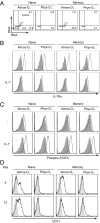
Fig. 1.
Fig. 1.
Physiological O2 levels diminish IL-7–induced cell cycle entry while maintaining proximal IL-7Rα signaling. Naive and memory CD4 T cell populations isolated from adult peripheral blood (APB) were stimulated with IL-7 (10 ng/mL) under 20% (Atmos-O2) or 2.5% (Phys-O2) O2 conditions. (A) Cell cycle entry was monitored as a function of DNA and RNA levels, using 7-amino-actinomycin-D (7AAD) and pyronin Y (PY), respectively. PY/7AAD cell cycle staining is shown at day 9, and the percentages of cells in the G1b (lower right quadrant) and S/G2/M stages (upper right quadrant) are indicated. Cells in the lower left quadrant are in G0/G1a. (B) Expression of CD127, the IL-7Rα chain, was monitored at day 3, and specific staining (open histograms) compared with control isotype (closed histograms) is shown. (C) STAT5 phosphorylation was measured using an anti–phospho-STAT5 polyclonal antibody. Specific (open histograms) relative to control isotype staining (closed histograms) is shown in the presence or absence of IL-7. (D) CD71 transferrin receptor expression (open histograms) and isotype controls (closed histograms) were monitored at days 9 and 12 of IL-7 stimulation. Data are representative of three independent experiments comprising three to six different donors.
Fig. 2.
Fig. 2.
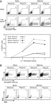
Permissivity of IL-7–stimulated CD4 T lymphocytes to infection by X4- and VSV-G–pseudotyped HIV-1 virions does not correlate with cell cycle kinetics under physiological O2 concentrations. (A) Naive and memory CD4 T cell populations isolated from APB were stimulated for 1, 3, 6, or 9 d with IL-7 (10 ng/mL) under 20% (Atmos-O2) or 2.5% (Phys-O2) conditions and then infected with an X4-tropic HIV-1 vector harboring the EGFP reporter gene. Single-round infected cells were detected by monitoring EGFP expression 48 h postinfection. Representative dot plots are shown for infections performed at day 9 of IL-7 stimulation, and the percentages of infected EGFP+ cells are indicated (Upper). The Lower panel shows a quantification of the percentages of EGFP+ cells from infections performed at the indicated time points. (B) Nine days post–IL-7 stimulation, naive and memory CD4 T cells were infected with a VSV-G envelope-pseudotyped HIV-1 vector expressing EGFP. The percentages of EGFP+ cells are indicated (Upper). The corresponding level of cell cycle entry, assessed at the time of infection by PY/7AAD staining, is shown (Lower). The percentages of cells in the G1b (lower right quadrant) and S/G2/M (upper right quadrant) phases of the cell cycle are indicated (Lower). Data are representative of three independent experiments comprising three to eight different donors.
Fig. 3.
Fig. 3.
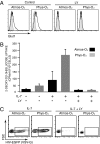
IL-7–induced expression and function of the Glut1 glucose transporter are enhanced under physiological O2 conditions, and its PI3K-mediated abrogation results in attenuated HIV-1 infection. (A) IL-7–stimulated APB CD4 T cells were cultured in the absence or presence of the PI3K inhibitor LY294002 (LY; 15 μM) and, at day 3, Glut1 expression was monitored. Representative histograms show specific Glut1 staining (open) relative to control staining (filled). (B) Glucose uptake was monitored in 1 × 10⁶ CD4 T cells cultured for 3 d in the absence (−) or presence (+) of IL-7 and the LY inhibitor, as above. Lymphocytes were incubated with 2-deoxy-d-[1-3H]glucose (2 μCi) for 10 min at room temperature. Results are expressed as mean cpm ± SD for triplicate sample analysis from cells cultured under 20% (black) or 2.5% (gray) O2 conditions. (C) IL-7–stimulated CD4 T cells, cultured in the absence or presence of LY, were infected with VSV-G–pseudotyped HIV-EGFP virions. Infection was monitored 48 h later and the percentages of EGFP+ cells are indicated. Data are representative of results obtained in six independent experiments comprising seven different donors.
Fig. 4.
Fig. 4.
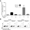
Inhibition of Glut1 up-regulation attenuates HIV-1 infection under atmospheric and physiological O2 conditions. (A) CD4 T cells from APB were transfected with Glut1-specific siRNAs (siGlut1) and then stimulated under 20% (Atmos-O2) or 2.5% (Phys-O2) conditions for 24 h. Glucose uptake was monitored by incubating cells with 2-deoxy-d-[1-3H]glucose (2 μCi) for 10 min at room temperature. Results are expressed as mean cpm ± SD for triplicate samples. (B) CD4 T cells stimulated following transfection with control (siCTRL) or Glut1-specific siRNAs were infected with VSV-G–pseudotyped HIV-EGFP virions. Infection was monitored at 48 h and the percentages of EGFP+ cells are indicated. Data are representative of results obtained in four independent experiments comprising four different donors.
Fig. 5.
Fig. 5.
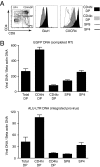
Glut1 expression is associated with increased permissivity of the CD4hiCXCR4hi DP human thymocyte subset to VSV-G–pseudotyped HIV-1 infection. (A) Glut1 and CXCR4 staining in the CD4hiDP, CD4loDP, SP4, and SP8 human thymocyte subsets are shown as histogram overlays. The gates used to define and FACS sort these subsets are shown, and the percentages of cells in each quadrant are indicated. (B) Freshly isolated human thymocytes were infected for 12 h with a single-round VSV-G–pseudotyped HIV-1. Thymocyte populations were subsequently sorted by FACS and quantitative PCR analysis was performed to detect EGFP DNA (Upper), indicative of completed reverse transcription, as well as a two-step nested PCR for Alu-LTR DNA to detect integrated provirus (Lower). Signals were normalized to β-actin DNA. Results for each subset are expressed as means ± SD of triplicate samples.

Similar articles

Cited by

References

    1. Eckstein DA, et al. HIV-1 actively replicates in naive CD4+ T cells residing within human lymphoid tissues. Immunity. 2001;15:671–682. - PubMed
    1. Kinter A, Moorthy A, Jackson R, Fauci AS. Productive HIV infection of resting CD4+ T cells: Role of lymphoid tissue microenvironment and effect of immunomodulating agents. AIDS Res Hum Retroviruses. 2003;19:847–856. - PubMed
    1. Korin YD, Zack JA. Progression to the G1b phase of the cell cycle is required for completion of human immunodeficiency virus type 1 reverse transcription in T cells. J Virol. 1998;72:3161–3168. - PMC - PubMed
    1. Biancotto A, et al. HIV-1 induced activation of CD4+ T cells creates new targets for HIV-1 infection in human lymphoid tissue ex vivo. Blood. 2008;111:699–704. - PMC - PubMed
    1. Vatakis DN, Nixon CC, Zack JA. Quiescent T cells and HIV: An unresolved relationship. Immunol Res. 2010;48(1–3):110–121. - PMC - PubMed

Publication types