. 2012 Feb 14;109(7):2595-600.
doi: 10.1073/pnas.1121604109. Epub 2012 Jan 30.

Chondroitinase ABC promotes selective reactivation of somatosensory cortex in squirrel monkeys after a cervical dorsal column lesion

Affiliations

Chondroitinase ABC promotes selective reactivation of somatosensory cortex in squirrel monkeys after a cervical dorsal column lesion

Charnese Bowes et al. Proc Natl Acad Sci U S A. .

Abstract

After large but incomplete lesions of ascending dorsal column afferents in the cervical spinal cord, the hand representation in the contralateral primary somatosensory cortex (area 3b) of monkeys is largely or completely unresponsive to touch on the hand. However, after weeks of spontaneous recovery, considerable reactivation of the hand territory in area 3b can occur. Because the reactivation process likely depends on the sprouting of remaining axons from the hand in the cuneate nucleus of the lower brainstem, we sought to influence cortical reactivation by treating the cuneate nucleus with an enzyme, chondroitinase ABC, that digests perineuronal nets, promoting axon sprouting. Dorsal column lesions were placed at a spinal cord level (C5/C6) that allowed a portion of ascending afferents from digit 1 to survive in squirrel monkeys. After 11-12 wk of recovery, the contralateral forelimb cortex was reactivated by stimulating digit 1 more extensively in treated monkeys than in control monkeys. The results are consistent with the proposal that the treatment enhances the sprouting of digit 1 afferents in the cuneate nucleus and that this sprouting allowed these preserved inputs to activate cortex more effectively.


Conflict of interest statement

The authors declare no conflict of interest.

Figures
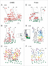
Fig. 1.
Fig. 1.
The organization of the forelimb region of the primary somatosensory cortex (area 3b) of treated (chABC) and control (P-ase) monkeys 11–12 wk after a unilateral lesion of the contralateral dorsal columns of the cervical spinal cord. The lesion was placed at the C5-C6 level to preserve some of the ascending afferents from D1 while removing most or all ascending afferents from digits 2–5 to the cuneate nucleus of the lower brainstem. At the time of the lesion, the squirrel monkey received either a brainstem injection of chABC or a control injection of P-ase. The injection was made just lateral to the cuneate nucleus to avoid damage but to digest the perineuronal net and promote axon sprouting. In each illustrated case, a surface view of somatosensory cortex indicates the locations of microelectrode penetrations where neurons responded to touch on the face (green dots), D1 (red dots), digits 2–5 (colors shown in Inset), or other parts of the palm and forearm (gray dots). Penetration sites where neurons failed to respond to touch are marked with Xs. The gray dotted line indicates cortical territory where D1 receptive fields (in red) were heavily represented. Note that the treated cases (AC) had more sites with neurons responsive to touch on D1 than did the cases with a control injection (DF). Inset shows the location of primary somatosensory cortex on a lateral view of a squirrel monkey brain. The view of somatosensory cortex is enlarged on the right, with the normal locations of the territories activated by touch on digits 1–5 and the face indicated by colors. Cortex caudal to the digit territories is activated from the palm; cortex medial to the digits is activated from the forearm. In A–F, cortex caudal to area 3b (S1) is area 1 followed by area 2 (1/2); cortex rostral to area 3b is area 3a and primary motor cortex (M1). Stars indicate locations where an electrolytic microlesion was placed for reference. The thin line across area 3b marks the location of a myelin-light septum that marks the face/hand boundary in normal monkeys and was identified in these monkeys histologically.
Fig. 2.
Fig. 2.
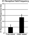
The proportion of recording sites responsive to touch on D1 to the total number of responsive sites in the forelimb area of 3b for the five control (P-ase) and five treated (chABC) cases. A significantly greater proportion of cortical territory was responsive to cutaneous stimulation of D1 in the chABC-treated group. n = 10. P < 0.05.
Fig. 3.
Fig. 3.
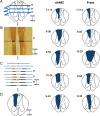
The extent of the spinal cord lesions in the five treated monkeys (chABC) and the five control monkeys (P-ase). (A) The lesions were reconstructed from a series of horizontal sections through the lesion site in the cervical spinal cord. Section numbers and the locations of the cuneate fasciculus (CF) and the gracile fasciculus (GF) are indicated. (B) A photomicrograph of section 37 stained for cytochrome oxidase. The white matter, gray matter (black), midline (gray), and lesion borders (yellow) were outlined for each section, and these lines were stacked as in C to produce D. The pinholes on the right in B were made in the intact spinal cord and later were used for realignment of each section. In both groups, the lesions of the cuneate fasciculus were complete or nearly complete.

Similar articles

Cited by

References

    1. Merzenich MM, et al. Progression of change following median nerve section in the cortical representation of the hand in areas 3b and 1 in adult owl and squirrel monkeys. Neuroscience. 1983;10:639–665. - PubMed
    1. Jain N, Catania KC, Kaas JH. Deactivation and reactivation of somatosensory cortex after dorsal spinal cord injury. Nature. 1997;386:495–498. - PubMed
    1. Darian-Smith C, Brown S. Functional changes at periphery and cortex following dorsal root lesions in adult monkeys. Nat Neurosci. 2000;3:476–481. - PubMed
    1. Jain N, Qi H-X, Collins CE, Kaas JH. Large-scale reorganization in the somatosensory cortex and thalamus after sensory loss in macaque monkeys. J Neurosci. 2008;28:11042–11060. - PMC - PubMed
    1. Qi HX, Chen LM, Kaas JH. Reorganization of somatosensory cortical areas 3b and 1 after unilateral section of dorsal columns of the spinal cord in squirrel monkeys. J Neurosci. 2011;31:13662–13675. - PMC - PubMed

Publication types

Substances