. 2012 Mar 27;6(3):2174-88.
doi: 10.1021/nn204102q. Epub 2012 Feb 14.

Delivery of small interfering RNA by peptide-targeted mesoporous silica nanoparticle-supported lipid bilayers

Affiliations

Delivery of small interfering RNA by peptide-targeted mesoporous silica nanoparticle-supported lipid bilayers

Carlee E Ashley et al. ACS Nano. .

Abstract

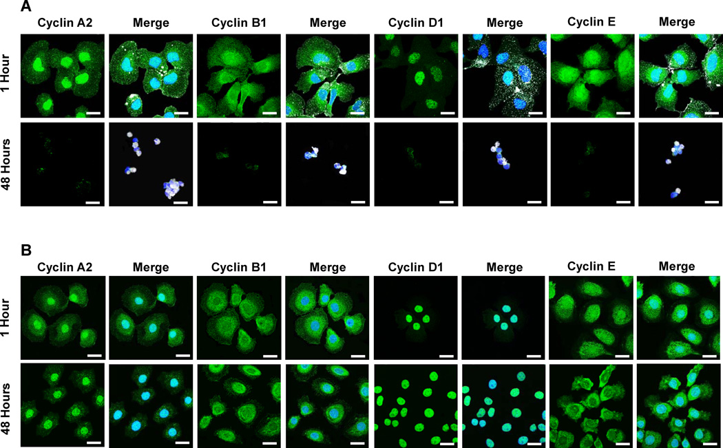
The therapeutic potential of small interfering RNAs (siRNAs) is severely limited by the availability of delivery platforms that protect siRNA from degradation, deliver it to the target cell with high specificity and efficiency, and promote its endosomal escape and cytosolic dispersion. Here we report that mesoporous silica nanoparticle-supported lipid bilayers (or "protocells") exhibit multiple properties that overcome many of the limitations of existing delivery platforms. Protocells have a 10- to 100-fold greater capacity for siRNA than corresponding lipid nanoparticles and are markedly more stable when incubated under physiological conditions. Protocells loaded with a cocktail of siRNAs bind to cells in a manner dependent on the presence of an appropriate targeting peptide and, through an endocytic pathway followed by endosomal disruption, promote delivery of the silencing nucleotides to the cytoplasm. The expression of each of the genes targeted by the siRNAs was shown to be repressed at the protein level, resulting in a potent induction of growth arrest and apoptosis. Incubation of control cells that lack expression of the antigen recognized by the targeting peptide with siRNA-loaded protocells induced neither repression of protein expression nor apoptosis, indicating the precise specificity of cytotoxic activity. In terms of loading capacity, targeting capabilities, and potency of action, protocells provide unique attributes as a delivery platform for therapeutic oligonucleotides.


Figures

Figure 1
Figure 1. Schematic depicting the process used to synthesize siRNA-loaded mesoporous silica nanoparticle-supported lipid bilayers (protocells)
To form protocells loaded with therapeutic RNA and targeted to hepatocellular carcinomas (HCC), mesoporous silica cores modified with an amine-containing silane (AEPTMS) were first soaked in a solution of small interfering RNA (siRNA). Liposomes composed of DOPC, DOPE, cholesterol, and 18:1 PEG-2000 PE (55:5:30:10 mass ratio) were then fused to siRNA-loaded cores. The resulting supported lipid bilayer (SLB) was modified with a targeting peptide (SP94) that binds to HCC and an endosomolytic peptide (H5WYG) that promotes endosomal/lysosomal escape of internalized protocells. Peptides, modified with glycine-glycine (GG) spacers and C-terminal cysteine residues, were conjugated to primary amines present in DOPE moieties via a heterobifunctional crosslinker (SM(PEG)24) with a 9.5-nm polyethylene glycol (PEG) spacer. The SP94 and H5WYG sequences reported by Lo et al. and Moore et al. are given in red.
Figure 2
Figure 2. Characterization of the mesoporous silica nanoparticles that form the protocell core
(A) Transmission electron microscopy (TEM) image of multimodal silica nanoparticles formed via the emulsion processing technique described by Carroll et al. Scale bar = 100 nm. The inset shows a scanning electron microscopy (SEM) image of a 5-µm multimodal silica particle, in which surface-accessible pores are visible; large particles were used to enhance resolution. Inset scale bar = 200 nm. (B) Dynamic light scattering (DLS) of multimodal silica nanoparticles after size-based separation. Resulting particles had an average diameter of ~165 nm. (C) Nitrogen sorption isotherm for size-separated multimodal silica nanoparticles. The presence of hysteresis is consistent with a network of larger pores interconnected by smaller pores. (D) A cumulative pore volume plot, calculated from the adsorption branch of the isotherm in (C) using the Barrett-Joyner-Halenda (BJH) model, demonstrates the presence of large (23–30 nm) pores and small (3–13 nm) pores.
Figure 3
Figure 3. Protocells have a high capacity for siRNA, the release of which is triggered by acidic pH
(A) The concentrations of siRNA that can be loaded within 1010 protocells and lipid nanoparticles (LNPs). Zeta potential values for unmodified and AEPTMS-modified silica cores in 0.5 X PBS (pH 7.4) are −32 mV and +12 mV, respectively. (B) and (C) The rates at which siRNA is released from DOPC protocells with AEPTMS-modified cores, DOPC LNPs, and DOTAP LNPs upon exposure to a pH 7.4 simulated body fluid (B) or a pH 5.0 buffer (C) at 37°C. The average diameters of siRNA-loaded protocells, DOPC LNPs, and DOTAP LNPs were 178-nm (± 24.3-nm), 135-nm (± 19.1-nm), and 144-nm (± 14.8-nm), respectively. Error bars represent 95% confidence intervals (1.96 σ) for n = 3.
Figure 4
Figure 4. siRNA-loaded, SP94-targeted protocells silence various cyclin family members in HCC but not hepatocytes
(A) and (B) Dose (A) and time (B) dependent decreases in the expression of cyclin A2, B1, D1, and E protein upon exposure of Hep3B to siRNA-loaded, SP94-targeted protocells. 1 × 10⁶ cells were continually exposed to various concentrations of siRNA for 48 hours in (A) and to 125 pM of siRNA for various periods of time in (B). Cyclin A2 mRNA levels are included for comparison. Protein concentrations were determined via flow cytometry analysis of cells stained by immunofluorescence. mRNA concentrations were determined by real time PCR. (C, left axis) Percentages of initial cyclin A2 protein concentrations that remain upon exposure of 1 × 10⁶ Hep3B or hepatocytes to 125 pM of siRNA, loaded within DOPC protocells or DOTAP lipid nanoparticles (LNPs), for 48 hours at 37°C. (C, right axis) The number of siRNA-loaded, SP94-targeted DOPC protocells or DOTAP LNPs that must be incubated with 1 × 10⁶ Hep3B cells to reduce expression of cyclin A2 protein to 10% of the initial concentration. DOPC LNPs were omitted from these experiments, as well as all subsequent analyses, as their efficacy was similar to that of free siRNA. Protocell SLBs were composed of DOPC with 5 wt% DOPE, 30 wt% cholesterol, and 10 wt% PEG-2000 and were modified with 0.015 wt% SP94 and 0.500 wt% H5WYG. DOTAP LNPs were prepared using a 55:5:30:10 ratio of DOTAP:DOPE:cholesterol:PEG-2000 PE and were modified with 0.015 wt% SP94, and 0.500 wt% H5WYG. Error bars represent 95% confidence intervals (1.96 σ) for n = 3.
Figure 5
Figure 5. Confocal fluorescence microscopy images of Hep3B (A) and hepatocytes (B) after exposure to siRNA-loaded, SP94-targeted protocells for 1 hour or 48 hours at 37°C
Cells were incubated with a 10-fold excess of Alexa Fluor 647-labeled protocells (white) prior to being fixed, permeablized, and stained with Hoechst 33342 (blue) and Alexa Fluor 488-labeled antibodies against cyclin A2, cyclin B1, cyclin D1, or cyclin E (green). Protocell SLBs were composed of DOPC with 5 wt% DOPE, 30 wt% cholesterol, and 10 wt% PEG-2000 and were modified with 0.015 wt% SP94 and 0.500 wt% H5WYG. Scale bars = 20 µm.
Figure 6
Figure 6. SP94-targeted protocells loaded with the cyclin-specific siRNA cocktail induce growth arrest and apoptosis in HCC without affecting hepatocyte viability
(A) – (C) The percentage of 1 × 10⁶ Hep3B that were proliferating (A), arrested in G0/G1 or G2/M (B), or apoptotic (C) upon exposure to SP94-targeted protocells loaded with 125 pM of the cyclin-specific siRNA cocktail for various periods of time at 37°C. Proliferation was determined using a flow cytometric assay for 5-bromo-2’-deoxyuridine (BrdU) incorporation, where cells positive for BrdU incorporation were considered to be actively proliferating. The numbers of cells in G0/G1, S, or G2/M phases of the cell cycle was determined via Hoechst 33342 staining followed by flow cytometry-based cell cycle analysis. Apoptosis was quantified using Alexa Fluor 488-labeled annexin V and propidium iodide (PI). Cells positive for annexin V were considered to be in the early stages of apoptosis, while cells positive for both annexin V and PI were considered to be in the late stages of apoptosis; the total number of apoptotic cells was determined by adding the numbers of cells in early and late apoptosis. (D, left axis) The percentages of 1 × 10⁶ Hep3B or heaptocytes that become apoptotic, i.e. double-positive for Alexa Fluor 488-labeled annexin V and propidium iodide, upon exposure to 125 pM of the siRNA cocktail, loaded within DOPC protocells or DOTAP lipid nanoparticles (LNPs), for 48 hours at 37°C. (D, right axis) The number of siRNA-loaded, SP94-targeted DOPC protocells or DOTAP LNPs that must be incubated with 1 × 10⁶ Hep3B to induce apoptosis in 90% of cells in the population. Protocell SLBs were composed of DOPC with 5 wt% DOPE, 30 wt% cholesterol, and 10 wt% PEG-2000 and were modified with 0.015 wt% SP94 and 0.500 wt% H5WYG. DOTAP LNPs were prepared using a 55:5:30:10 ratio of DOTAP:DOPE:cholesterol:PEG-2000 PE and were modified with 0.015 wt% SP94, and 0.500 wt% H5WYG. Error bars represent 95% confidence intervals (1.96 σ) for n = 3.
Figure 7
Figure 7. Confocal fluorescence microscopy images of Hep3B (A) and hepatocytes (B) after exposure to siRNA-loaded, SP94-targeted protocells for 1 hour or 48 hours at 37°C
Cells were incubated with a 10-fold excess of Alexa Fluor 647-labeled protocells (white) prior to being stained with Hoechst 33342 (blue), Alexa Fluor 488-labeled annexin V (green), and propidium iodide (red). Differential Interference Contrast (DIC) images are included to show cell morphology. Protocell SLBs were composed of DOPC with 5 wt% DOPE, 30 wt% cholesterol, and 10 wt% PEG-2000 and were modified with 0.015 wt% SP94 and 0.500 wt% H5WYG. Scale bars = 20 µm.

Similar articles

Cited by

References

    1. Peer D, Karp JM, Hong S, Farokhzad OC, Margalit R, Langer R. Nanocarriers as an Emerging Platform for Cancer Therapy. Nat Nano. 2007;2:751–760. - PubMed
    1. Petros RA, DeSimone JM. Strategies in the Design of Nanoparticles for Therapeutic Applications. Nat. Rev. Drug Discov. 2010;9:615–627. - PubMed
    1. Wang M, Thanou M. Targeting Nanoparticles to Cancer. Pharm. Res. 2010;62:90–99. - PubMed
    1. Meister G, Tuschl T. Mechanisms of Gene Silencing by Double-Stranded RNA. Nature. 2004;431:343–349. - PubMed
    1. Rana TM. Illuminating the Silence: Understanding the Structure and Function of Small RNAs. Nat. Rev. Mol. Cell Biol. 2007;8:23–36. - PubMed

Publication types