

Aggregatibacter actinomycetemcomitans leukotoxin cytotoxicity occurs through bilayer destabilization
- PMID: 22309134
- PMCID: PMC3412409
- DOI: 10.1111/j.1462-5822.2012.01762.x
Aggregatibacter actinomycetemcomitans leukotoxin cytotoxicity occurs through bilayer destabilization
Abstract
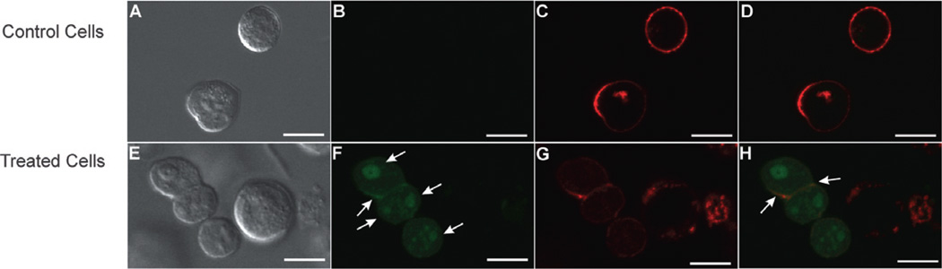
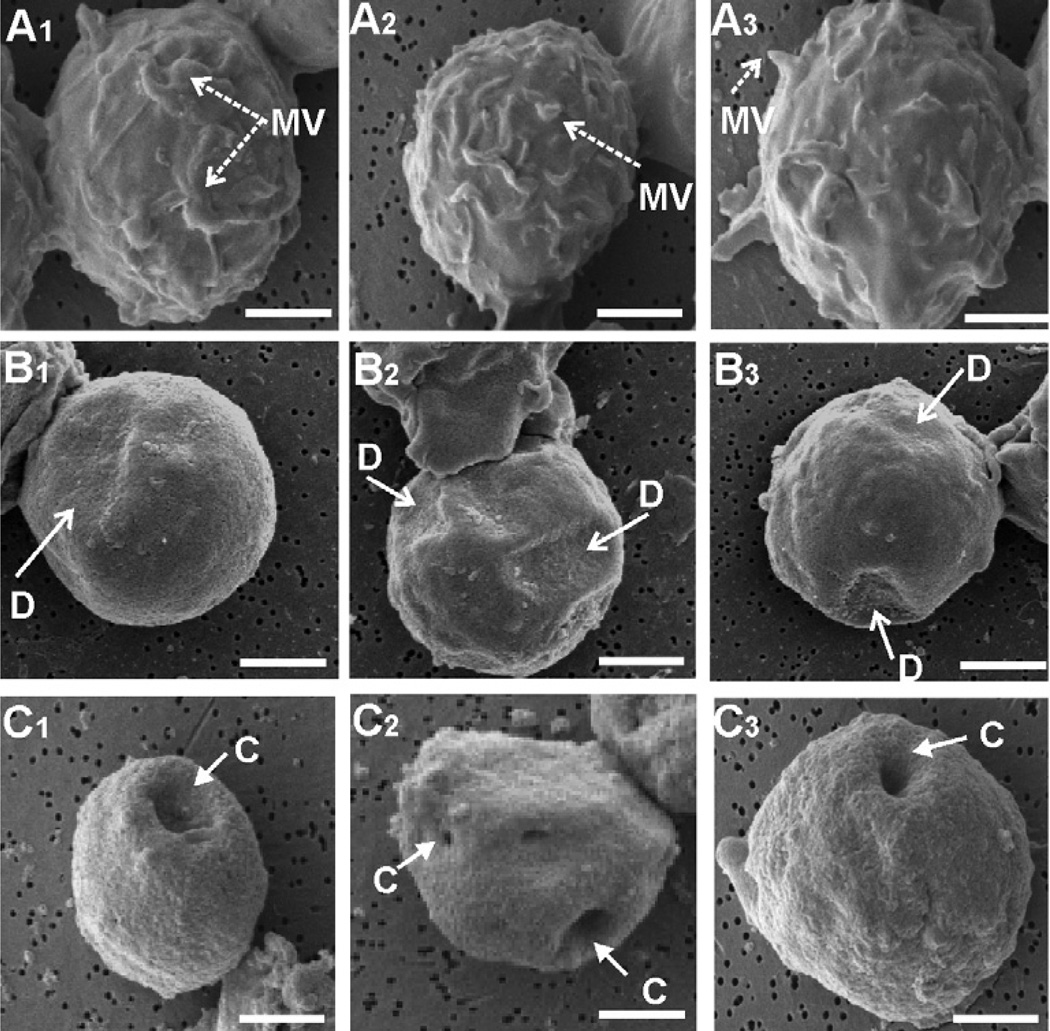
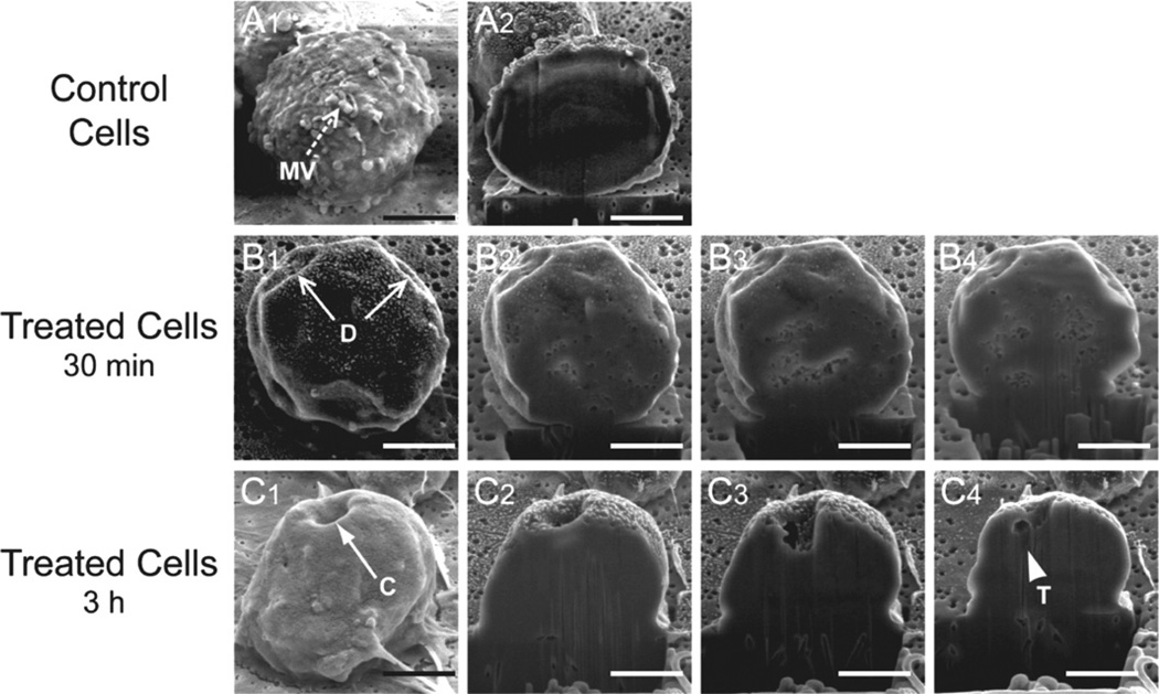
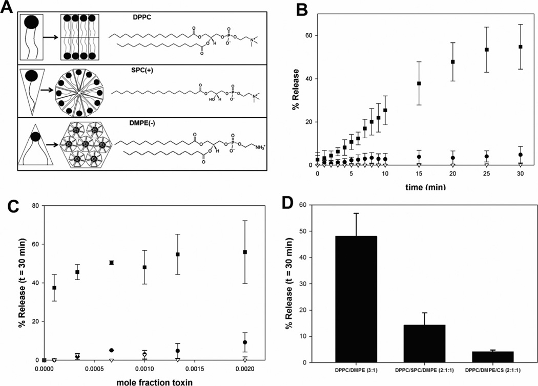
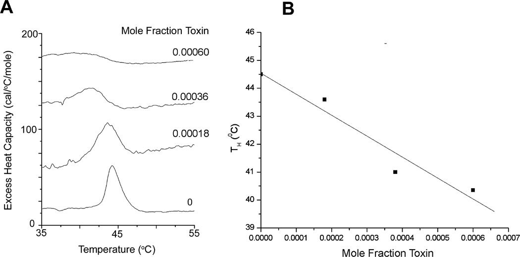
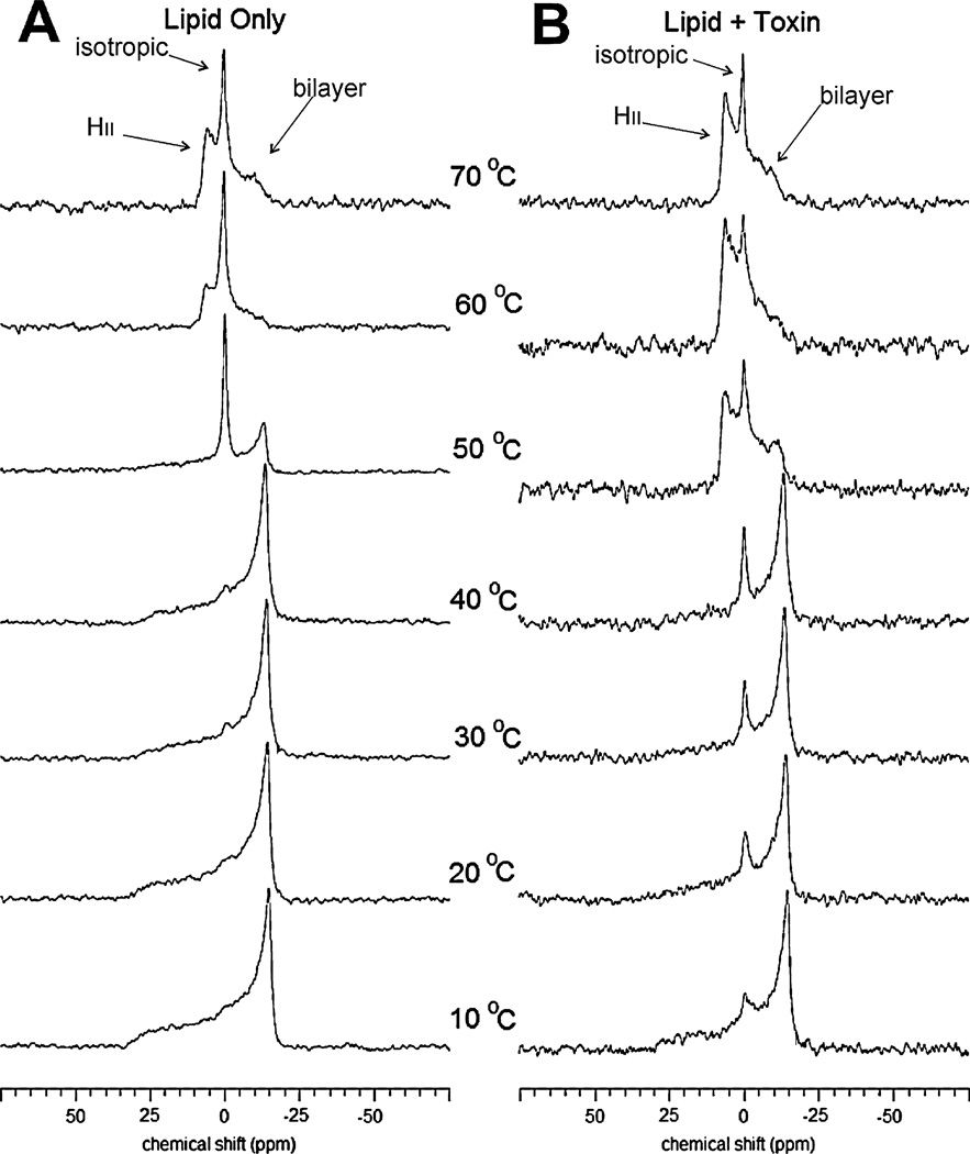
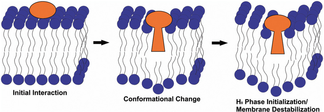
The Gram-negative bacterium, Aggregatibacter actinomycetemcomitans, is a common inhabitant of the human upper aerodigestive tract. The organism produces an RTX (Repeats in ToXin) toxin (LtxA) that kills human white blood cells. LtxA is believed to be a membrane-damaging toxin, but details of the cell surface interaction for this and several other RTX toxins have yet to be elucidated. Initial morphological studies suggested that LtxA was bending the target cell membrane. Because the ability of a membrane to bend is a function of its lipid composition, we assessed the proficiency of LtxA to release of a fluorescent dye from a panel of liposomes composed of various lipids. Liposomes composed of lipids that form nonlamellar phases were susceptible to LtxA-induced damage while liposomes composed of lipids that do not form non-bilayer structures were not. Differential scanning calorimetry demonstrated that the toxin decreased the temperature at which the lipid transitions from a bilayer to a nonlamellar phase, while (31) P nuclear magnetic resonance studies showed that the LtxA-induced transition from a bilayer to an inverted hexagonal phase occurs through the formation of an isotropic intermediate phase. These results indicate that LtxA cytotoxicity occurs through a process of membrane destabilization.
© 2012 Blackwell Publishing Ltd.
Figures
Similar articles
-
Aggregatibacter actinomycetemcomitans leukotoxin utilizes a cholesterol recognition/amino acid consensus site for membrane association.J Biol Chem. 2013 Aug 9;288(32):23607-21. doi: 10.1074/jbc.M113.486654. Epub 2013 Jun 21. J Biol Chem. 2013. PMID: 23792963 Free PMC article.
-
Toxin-triggered liposomes for the controlled release of antibiotics to treat infections associated with the gram-negative bacterium, Aggregatibacter actinomycetemcomitans.Colloids Surf B Biointerfaces. 2024 Jun;238:113870. doi: 10.1016/j.colsurfb.2024.113870. Epub 2024 Mar 21. Colloids Surf B Biointerfaces. 2024. PMID: 38555763 Free PMC article.
-
Membrane association and destabilization by Aggregatibacter actinomycetemcomitans leukotoxin requires changes in secondary structures.Mol Oral Microbiol. 2013 Oct;28(5):342-53. doi: 10.1111/omi.12028. Epub 2013 May 16. Mol Oral Microbiol. 2013. PMID: 23678967 Free PMC article.
-
Aggregatibacter actinomycetemcomitans leukotoxin: from threat to therapy.J Dent Res. 2010 Jun;89(6):561-70. doi: 10.1177/0022034510363682. Epub 2010 Mar 3. J Dent Res. 2010. PMID: 20200418 Free PMC article. Review.
-
Aggregatibacter actinomycetemcomitans Leukotoxin (LtxA; Leukothera®): Mechanisms of Action and Therapeutic Applications.Toxins (Basel). 2019 Aug 26;11(9):489. doi: 10.3390/toxins11090489. Toxins (Basel). 2019. PMID: 31454891 Free PMC article. Review.
Cited by
-
Kingella kingae RtxA Cytotoxin in the Context of Other RTX Toxins.Microorganisms. 2022 Feb 27;10(3):518. doi: 10.3390/microorganisms10030518. Microorganisms. 2022. PMID: 35336094 Free PMC article. Review.
-
Aggregatibacter actinomycetemcomitans leukotoxin: From mechanism to targeted anti-toxin therapeutics.Mol Oral Microbiol. 2020 Jun;35(3):85-105. doi: 10.1111/omi.12284. Epub 2020 Mar 10. Mol Oral Microbiol. 2020. PMID: 32061022 Free PMC article. Review.
-
Inhibition of LtxA toxicity by blocking cholesterol binding with peptides.Mol Oral Microbiol. 2016 Feb;31(1):94-105. doi: 10.1111/omi.12133. Epub 2015 Oct 12. Mol Oral Microbiol. 2016. PMID: 26352738 Free PMC article.
-
Aggregatibacter actinomycetemcomitans Leukotoxin Is Delivered to Host Cells in an LFA-1-Indepdendent Manner When Associated with Outer Membrane Vesicles.Toxins (Basel). 2018 Oct 13;10(10):414. doi: 10.3390/toxins10100414. Toxins (Basel). 2018. PMID: 30322160 Free PMC article.
-
Membrane Interaction Characteristics of the RTX Toxins and the Cholesterol-Dependence of Their Cytolytic/Cytotoxic Activity.Int J Mol Sci. 2024 Mar 8;25(6):3131. doi: 10.3390/ijms25063131. Int J Mol Sci. 2024. PMID: 38542105 Free PMC article. Review.
References
-
- Alonso A, Goni FM, Buckley JT. Lipids favoring inverted phase enhance the ability of aerolysin to permeabilize liposome bilayers. Biochemistry. 2000;39:14019–14024. - PubMed
-
- Anderluh G, Serra MD, Viero G, Guella G, Macek P, Menestrina G. Pore formation by Equinatoxin II, a eukaryotic protein toxin, occurs by induction of non-lamellar lipid structures. J Biol Chem. 2003;278:45216–45233. - PubMed
-
- Balakrishnan L, Hughes C, Koronakis V. Substrate-triggered recruitment of the TolC channel-tunnel during type I export of hemolysin by Escherichia coli. J Mol Biol. 2001;313:501–510. - PubMed
MeSH terms
Substances
Grants and funding
LinkOut - more resources
Full Text Sources
