. 2012 Apr 6;11(4):2508-20.
doi: 10.1021/pr201206w. Epub 2012 Mar 13.

Lectin chromatography/mass spectrometry discovery workflow identifies putative biomarkers of aggressive breast cancers

Affiliations

Lectin chromatography/mass spectrometry discovery workflow identifies putative biomarkers of aggressive breast cancers

Penelope M Drake et al. J Proteome Res. .

Abstract

We used a lectin chromatography/MS-based approach to screen conditioned medium from a panel of luminal (less aggressive) and triple negative (more aggressive) breast cancer cell lines (n=5/subtype). The samples were fractionated using the lectins Aleuria aurantia (AAL) and Sambucus nigra agglutinin (SNA), which recognize fucose and sialic acid, respectively. The bound fractions were enzymatically N-deglycosylated and analyzed by LC-MS/MS. In total, we identified 533 glycoproteins, ∼90% of which were components of the cell surface or extracellular matrix. We observed 1011 glycosites, 100 of which were solely detected in ≥3 triple negative lines. Statistical analyses suggested that a number of these glycosites were triple negative-specific and thus potential biomarkers for this tumor subtype. An analysis of RNaseq data revealed that approximately half of the mRNAs encoding the protein scaffolds that carried potential biomarker glycosites were up-regulated in triple negative vs luminal cell lines, and that a number of genes encoding fucosyl- or sialyltransferases were differentially expressed between the two subtypes, suggesting that alterations in glycosylation may also drive candidate identification. Notably, the glycoproteins from which these putative biomarker candidates were derived are involved in cancer-related processes. Thus, they may represent novel therapeutic targets for this aggressive tumor subtype.


Figures

Fig. 1
Fig. 1. Breast cancer cell lines have a complex repertoire of SNA-reactive glycoproteins and exhibit cell surface staining with this lectin
(A) Lysates from a panel of 8 breast cancer cell lines, which included triple negative (1–6) and luminal (7, 8) subtypes, were electrophoretically separated, transferred to nitrocellulose, and probed with SNA. Lane 1. MDAMB468, 2. HCC38, 3. HCC1500, 4. HS578T, 5. MDAMB157, 6. MDAMB231, 7. T47D, 8. UCC812. (B) Non-permeabilized HS578T cells were stained with fluorescein-conjugated SNA and imaged by fluorescence microscopy (magnification 60x).
Fig. 2
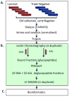
Fig. 2. The experimental workflow
(A) CM samples from breast cancer cell lines established from five luminal and 5 triple negative tumors were prepared in one laboratory, then distributed to 3 sites. (B) Each group separated the 10 CM samples, in duplicate, by AAL or SNA chromatography, which generated 40 fractions. The samples were deglycosylated using PNGaseF and analyzed in duplicate by LC-MS/MS, yielding a total of 80 MS/MS data sets per site. (C) Files were transferred to a central location for bioinformatic analyses.
Fig. 3
Fig. 3. Diagrammatic summary of the glycosite (glycoprotein) enrichment data according to lectin type (AAL vs. SNA) and CM samples (luminal vs. triple negative) showed distinct and overlapping specificities
(A) The intersecting circles depict the total number of N-glycosites (glycoproteins) captured by each lectin. (B and C) Venn diagrams illustrating the chromatographic separation of luminal (LUM) and triple negative (TN) CM samples.
Fig. 4
Fig. 4. Lectin capture resulted in significant glycopeptide enrichment
The percent enrichment for the separations performed using AAL (left) or SNA (right) at Sites M (top), X (middle), and S (bottom). The dark line indicates the median; the box depicts the first and third quartiles; the whiskers show the minimum and maximum values observed. Sites M and X acquired data using QSTAR Elite instruments, while Site S used an Orbitrap mass spectrometer.
Fig. 5
Fig. 5. Nearly 90% of identified glycoproteins resided in the plasma membrane or extracellular compartments
A portion (241/560) of the identified glycoproteins were annotated in the cellular component of Gene Ontology. Of these, the great majority were cell surface or secreted molecules.
Fig. 6
Fig. 6. Putative triple negative-specific glycosites (glycoproteins) enriched by AAL or SNA
The criteria applied were detection in ≥3 triple negative and 0 luminal cell line CMs.

Similar articles

Cited by

References

    1. Drake PM, Cho W, Li B, Prakobphol A, Johansen E, Anderson NL, Regnier FE, Gibson BW, Fisher SJ. Sweetening the Pot: Adding Glycosylation to the Biomarker Discovery Equation. Clin Chem. 2010;56:223–236. - PMC - PubMed
    1. Hart GW, Copeland RJ. Glycomics hits the big time. Cell. 2010;143(5):672–6. - PMC - PubMed
    1. Clowers BH, Dodds ED, Seipert RR, Lebrilla CB. Site determination of protein glycosylation based on digestion with immobilized nonspecific proteases and Fourier transform ion cyclotron resonance mass spectrometry. J Proteome Res. 2007;6(10):4032–40. - PubMed
    1. Duffy MJ, Evoy D, McDermott EW. CA 15–3: uses and limitation as a biomarker for breast cancer. Clin Chim Acta. 2010;411(23–24):1869–74. - PubMed
    1. Orntoft TF, Vestergaard EM. Clinical aspects of altered glycosylation of glycoproteins in cancer. Electrophoresis. 1999;20(2):362–71. - PubMed

Publication types

MeSH terms