Cardiovascular magnetic resonance by non contrast T1-mapping allows assessment of severity of injury in acute myocardial infarction
- PMID: 22309452
- PMCID: PMC3312869
- DOI: 10.1186/1532-429X-14-15
Cardiovascular magnetic resonance by non contrast T1-mapping allows assessment of severity of injury in acute myocardial infarction
Abstract
Background: Current cardiovascular magnetic resonance (CMR) methods, such as late gadolinium enhancement (LGE) and oedema imaging (T2W) used to depict myocardial ischemia, have limitations. Novel quantitative T1-mapping techniques have the potential to further characterize the components of ischemic injury. In patients with myocardial infarction (MI) we sought to investigate whether state-of the art pre-contrast T1-mapping (1) detects acute myocardial injury, (2) allows for quantification of the severity of damage when compared to standard techniques such as LGE and T2W, and (3) has the ability to predict long term functional recovery.
Methods: 3T CMR including T2W, T1-mapping and LGE was performed in 41 patients [of these, 78% were ST elevation MI (STEMI)] with acute MI at 12-48 hour after chest pain onset and at 6 months (6M). Patients with STEMI underwent primary PCI prior to CMR. Assessment of acute regional wall motion abnormalities, acute segmental damaged fraction by T2W and LGE and mean segmental T1 values was performed on matching short axis slices. LGE and improvement in regional wall motion at 6M were also obtained.
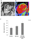
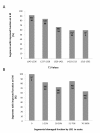
Results: We found that the variability of T1 measurements was significantly lower compared to T2W and that, while the diagnostic performance of acute T1-mapping for detecting myocardial injury was at least as good as that of T2W-CMR in STEMI patients, it was superior to T2W imaging in NSTEMI. There was a significant relationship between the segmental damaged fraction assessed by either by LGE or T2W, and mean segmental T1 values (P < 0.01). The index of salvaged myocardium derived by acute T1-mapping and 6M LGE was not different to the one derived from T2W (P = 0.88). Furthermore, the likelihood of improvement of segmental function at 6M decreased progressively as acute T1 values increased (P < 0.0004).
Conclusions: In acute MI, pre-contrast T1-mapping allows assessment of the extent of myocardial damage. T1-mapping might become an important complementary technique to LGE and T2W for identification of reversible myocardial injury and prediction of functional recovery in acute MI.
Figures


Similar articles
-
Combined T1-mapping and tissue tracking analysis predicts severity of ischemic injury following acute STEMI-an Oxford Acute Myocardial Infarction (OxAMI) study.Int J Cardiovasc Imaging. 2019 Jul;35(7):1297-1308. doi: 10.1007/s10554-019-01542-8. Epub 2019 Feb 16. Int J Cardiovasc Imaging. 2019. PMID: 30778713 Free PMC article.
-
T(1) mapping for the diagnosis of acute myocarditis using CMR: comparison to T2-weighted and late gadolinium enhanced imaging.JACC Cardiovasc Imaging. 2013 Oct;6(10):1048-1058. doi: 10.1016/j.jcmg.2013.03.008. Epub 2013 Sep 4. JACC Cardiovasc Imaging. 2013. PMID: 24011774
-
Native T1-mapping detects the location, extent and patterns of acute myocarditis without the need for gadolinium contrast agents.J Cardiovasc Magn Reson. 2014 May 23;16(1):36. doi: 10.1186/1532-429X-16-36. J Cardiovasc Magn Reson. 2014. PMID: 24886708 Free PMC article.
-
Tissue characterization of the myocardium: state of the art characterization by magnetic resonance and computed tomography imaging.Radiol Clin North Am. 2015 Mar;53(2):413-23. doi: 10.1016/j.rcl.2014.11.005. Epub 2014 Dec 18. Radiol Clin North Am. 2015. PMID: 25727003 Free PMC article. Review.
-
Role of cardiac T1 mapping and extracellular volume in the assessment of myocardial infarction.Anatol J Cardiol. 2018 Jun;19(6):404-411. doi: 10.14744/AnatolJCardiol.2018.39586. Epub 2018 Apr 10. Anatol J Cardiol. 2018. PMID: 29638222 Free PMC article. Review.
Cited by
-
Current T₁ and T₂ mapping techniques applied with simple thresholds cannot discriminate acute from chronic myocadial infarction on an individual patient basis: a pilot study.BMC Med Imaging. 2016 Apr 29;16:35. doi: 10.1186/s12880-016-0135-y. BMC Med Imaging. 2016. PMID: 27129879 Free PMC article.
-
Free-breathing three-dimensional simultaneous myocardial T1 and T2 mapping based on multi-parametric SAturation-recovery and Variable-flip-Angle.J Cardiovasc Magn Reson. 2024 Winter;26(2):101065. doi: 10.1016/j.jocmr.2024.101065. Epub 2024 Jul 24. J Cardiovasc Magn Reson. 2024. PMID: 39059610 Free PMC article.
-
Cardiovascular Magnetic Resonance Before Invasive Coronary Angiography in Suspected Non-ST-Segment Elevation Myocardial Infarction.JACC Cardiovasc Imaging. 2024 Sep;17(9):1044-1058. doi: 10.1016/j.jcmg.2024.05.007. Epub 2024 Jun 4. JACC Cardiovasc Imaging. 2024. PMID: 38970595 Free PMC article.
-
Relationship between native papillary muscle T1 time and severity of functional mitral regurgitation in patients with non-ischemic dilated cardiomyopathy.J Cardiovasc Magn Reson. 2016 Nov 16;18(1):79. doi: 10.1186/s12968-016-0301-y. J Cardiovasc Magn Reson. 2016. PMID: 27846845 Free PMC article.
-
Free-Breathing Ungated Radial Simultaneous Multi-Slice Cardiac T1 Mapping.J Magn Reson Imaging. 2025 Jun;61(6):2587-2600. doi: 10.1002/jmri.29676. Epub 2024 Dec 11. J Magn Reson Imaging. 2025. PMID: 39661447 Free PMC article.
References
-
- Lima JAC, Judd RM, Bazille A, Schulman SP, Atalar E, Zerhouni EA. Regional Heterogeneity of Human Myocardial Infarcts Demonstrated by Contrast-Enhanced MRI: Potential Mechanisms. Circulation. 1995;92:1117–1125. - PubMed
-
- Aletras AH, Tilak GS, Natanzon A, Hsu L-Y, Gonzalez FM, Hoyt RF, Arai AE. Retrospective Determination of the Area at Risk for Reperfused Acute Myocardial Infarction With T2-Weighted Cardiac Magnetic Resonance Imaging: Histopathological and Displacement Encoding With Stimulated Echoes (DENSE) Functional Validations. Circulation. 2006;113:1865–1870. doi: 10.1161/CIRCULATIONAHA.105.576025. - DOI - PubMed
Publication types
MeSH terms
Substances
Grants and funding
LinkOut - more resources
Full Text Sources
Medical
Miscellaneous

