. 2012 Apr 1;318(6):716-22.
doi: 10.1016/j.yexcr.2012.01.020. Epub 2012 Jan 28.

Epithelial cells utilize cortical actin/myosin to activate latent TGF-β through integrin α(v)β(6)-dependent physical force

Affiliations

Epithelial cells utilize cortical actin/myosin to activate latent TGF-β through integrin α(v)β(6)-dependent physical force

Marilyn M Giacomini et al. Exp Cell Res. .

Abstract

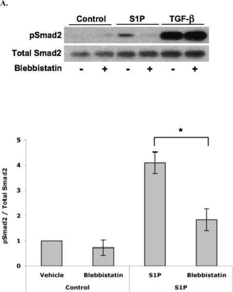
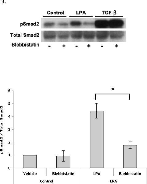
Transforming Growth Factor Beta (TGF-β) is involved in regulating many biological processes and disease states. Cells secrete cytokine as a latent complex that must be activated for it to exert its biological functions. We previously discovered that the epithelial-restricted integrin α(v)β(6) activates TGF-β and that this process is important in a number of in vivo models of disease. Here, we show that agonists of G-protein coupled receptors (Sphingosine-1-Phosphate and Lysophosphatidic Acid) which are ligated under conditions of epithelial injury directly stimulate primary airway epithelial cells to activate latent TGF-β through a pathway that involves Rho Kinase, non-muscle myosin, the α(v)β(6) integrin, and the generation of mechanical tension. Interestingly, lung epithelial cells appear to exert force on latent TGF-β using sub-cortical actin/myosin rather than the stress fibers utilized by fibroblasts and other traditionally "contractile" cells. These findings extend recent evidence suggesting TGF-β can be activated by integrin-mediated mechanical force and suggest that this mechanism is important for an integrin (α(v)β(6)) and a cell type (epithelial cells) that have important roles in biologically relevant TGF-β activation in vivo.


Figures

fig. 1
fig. 1. S1P induces αvβ6-mediated TGF-β activation via Rho Kinase
(A) Western blot for pSmad2 or Total Smad2 from lysates of NHBE cells treated with methanol (control) or S1P (1–30 μM) in the presence or absence of an anti-β6 antibody (40 μg/mL). (B) Western blots, as above, of lysates from NHBE cells treated with methanol (control), S1P (30 μM), and active TGF-β1 (15 pg/mL) in the presence or absence of an anti-β6 antibody (40 μg/mL), Rho Kinase inhibitor (Y-27632, 100 μM), and anti-TGF-β antibody (40 μg/mL). (C) Western blots of lysates of air-liquid interface cultures of NHBE cells treated with methanol (control), S1P (30 μM), and active TGF-β1 (15 pg/mL) in the presence or absence of an anti-β6 antibody (40 μg/mL), Y-27632 (100 μM), and anti-TGF-β antibody (40 μg/mL). *P < 0.05 for indicated comparison or versus the corresponding dose groups. The ratio of pSmad2 density to that of Total Smad2 was analyzed. All results are representative of at least three independent experiments. Error bars represent the standard error of the mean (s.e.m.).
fig. 2
fig. 2. S1P and LPA induced αvβ6-mediated TGF-β activation requires cellular tension
(A) Western blots for pSmad2 and Total Smad2 of lysates of NHBE cells treated with methanol (control), S1P (30 μM), and active TGF-β1 (15 pg/mL) in the presence or absence of Blebbistatin (20 μM). (B) Western blots, as above, of lysates of NHBE cells treated with water (control), LPA (50 μM), and active TGF-β1 (15 pg/mL) in the presence or absence of Blebbistatin (20 μM). (C) Western blots of lysates from NHBE cells cultured on various rigidities (0.025%, 0.05%, 0.1%, 0.2%) of polyacrylamide flexible substrates and treated with methanol (control), S1P (30 μM), and active TGF-β1 (15 pg/mL) in the presence or absence of an anti-β6 antibody (40 μg/mL) and anti-TGF-β antibody (40 μg/mL). *P < 0.05 for indicated comparison or versus the corresponding vehicle treated group. The ratio of pSmad2 density to that of Total Smad2 was analyzed. All results are representative of at least three independent experiments. Error bars represent the s.e.m.
fig. 3
fig. 3. S1P and LPA induced β6-mediated TGF-β activation results in cortical actin formation
(A) Phalloidin staining of the actin cytoskeleton in NHBE cells treated with methanol (vehicle) or S1P (30 μM) in the presence or absence of Y-27632 (100 μM) and Blebbistatin (20 μM). (B) Phalloidin staining, as above, in NHBE cells treated with water (vehicle) or LPA (50 μM) in the presence or absence of Y-27632 (100 μM) and Blebbistatin (20 μM). Arrows indicate cortical actin structures. Scale bars: 20 μm

Similar articles

Cited by

  • Generation and characterization of a diabody targeting the αvβ6 integrin.
    Kogelberg H, Miranda E, Burnet J, Ellison D, Tolner B, Foster J, Picón C, Thomas GJ, Meyer T, Marshall JF, Mather SJ, Chester K. Kogelberg H, et al. PLoS One. 2013 Sep 4;8(9):e73260. doi: 10.1371/journal.pone.0073260. eCollection 2013. PLoS One. 2013. PMID: 24023846 Free PMC article.
  • TGF-β activation and function in immunity.
    Travis MA, Sheppard D. Travis MA, et al. Annu Rev Immunol. 2014;32:51-82. doi: 10.1146/annurev-immunol-032713-120257. Epub 2013 Dec 2. Annu Rev Immunol. 2014. PMID: 24313777 Free PMC article. Review.
  • Why Stress Matters: An Introduction.
    Tschumperlin DJ. Tschumperlin DJ. Methods Mol Biol. 2021;2299:159-169. doi: 10.1007/978-1-0716-1382-5_12. Methods Mol Biol. 2021. PMID: 34028742 Review.
  • Matrix, mesenchyme, and mechanotransduction.
    Tschumperlin DJ. Tschumperlin DJ. Ann Am Thorac Soc. 2015 Mar;12 Suppl 1(Suppl 1):S24-9. doi: 10.1513/AnnalsATS.201407-320MG. Ann Am Thorac Soc. 2015. PMID: 25830830 Free PMC article. Review.
  • ROCKing pulmonary fibrosis.
    Sheppard D. Sheppard D. J Clin Invest. 2013 Mar;123(3):1005-6. doi: 10.1172/JCI68417. Epub 2013 Feb 22. J Clin Invest. 2013. PMID: 23434586 Free PMC article.

References

    1. Munger JS, Huang X, Kawakatsu H, Griffiths MJ, Dalton SL, Wu J, Pittet JF, Kaminski N, Garat C, Matthay MA, Rifkin DB, Sheppard D. The integrin alpha v beta 6 binds and activates latent TGF beta 1: a mechanism for regulating pulmonary inflammation and fibrosis. Cell. 1999;96:319–328. - PubMed
    1. Puthawala K, Hadjiangelis N, Jacoby SC, Bayongan E, Zhao Z, Yang Z, Devitt ML, Horan GS, Weinreb PH, Lukashev ME, Violette SM, Grant KS, Colarossi C, Formenti SC, Munger JS. Inhibition of integrin alpha(v)beta6, an activator of latent transforming growth factor-beta, prevents radiation-induced lung fibrosis. Am J Respir Crit Care Med. 2008;177:82–90. - PMC - PubMed
    1. Ma LJ, Yang H, Gaspert A, Carlesso G, Barty MM, Davidson JM, Sheppard D, Fogo AB. Transforming growth factor-beta-dependent and - independent pathways of induction of tubulointerstitial fibrosis in beta6(−/−) mice. Am J Pathol. 2003;163:1261–1273. - PMC - PubMed
    1. Hahm K, Lukashev ME, Luo Y, Yang WJ, Dolinski BM, Weinreb PH, Simon KJ, Chun Wang L, Leone DR, Lobb RR, McCrann DJ, Allaire NE, Horan GS, Fogo A, Kalluri R, Shield CF, 3rd, Sheppard D, Gardner HA, Violette SM. Alphav beta6 integrin regulates renal fibrosis and inflammation in Alport mouse. Am J Pathol. 2007;170:110–125. - PMC - PubMed
    1. Wang B, Dolinski BM, Kikuchi N, Leone DR, Peters MG, Weinreb PH, Violette SM, Bissell DM. Role of alphavbeta6 integrin in acute biliary fibrosis. Hepatology. 2007;46:1404–1412. - PMC - PubMed

Publication types