Comparative Study
. 2012 Mar;12(2):328-35.
doi: 10.1016/j.mito.2012.01.001. Epub 2012 Jan 28.

Microscale oxygraphy reveals OXPHOS impairment in MRC mutant cells

Affiliations
Comparative Study

Microscale oxygraphy reveals OXPHOS impairment in MRC mutant cells

F Invernizzi et al. Mitochondrion. 2012 Mar.

Abstract

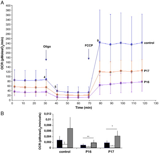
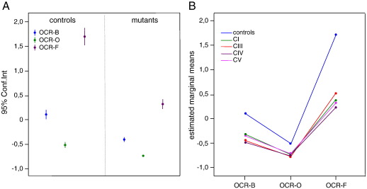
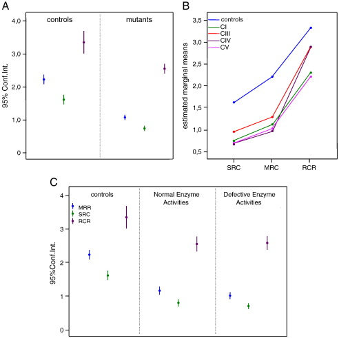
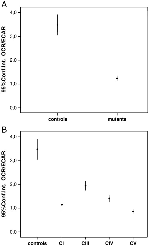
Given the complexity of the respiratory chain structure, assembly and regulation, the diagnostic workout for the identification of defects of oxidative phosphorylation (OXPHOS) is a major challenge. Spectrophotometric assays, that measure the activity of individual respiratory complexes in tissue and cell homogenates or isolated mitochondria, are highly specific, but their utilization is limited by the availability of sufficient biological material and intrinsic sensitivity. A further limitation is tissue specificity, which usually determines attenuation, or disappearance, in cultured fibroblasts, of defects detected in muscle or liver. We used numerous fibroblast cell lines derived from patients with OXPHOS deficiencies to set up experimental protocols required for the direct readout of cellular respiration using the Seahorse XF96 apparatus, which measures oxygen consumption rate (OCR) and extra-cellular acidification rate (ECAR) in 96 well plates. Results demonstrate that first level screening based on microscale oxygraphy is more sensitive, cheaper and rapid than spectrophotometry for the biochemical evaluation of cells from patients with suspected mitochondrial disorders.


Figures

Fig. 1
Fig. 1
OCR profile in control and mutant cell lines. A: OCR Traces, expressed as pMolesO2/min in control and mutant cell lines P16 and P17. Vertical lines indicate the time of addition of Oligomycin and FCCP. Values at points 4, 5, 9 reflect OCR-B, -O, F, respectively. B: OCR normalization to cell number. Black, white and gray histograms indicate OCR-B, -O, -F, respectively. Bars indicate the standard deviation (SD). * p < 0.05; ** p < 0.01 (unpaired, two-tail Student's t test).
Fig. 2
Fig. 2
Analysis of OCR-B, -O, -F. The z score values (dots) for OCR-B, -O, -F are shown for control vs. mutant cells. In panel A, the control and mutant cells were considered as single groups and expressed as the means (dots) and 95% confidence intervals, Conf.Int. (bars). ANOVA test for controls vs. mutants: OCR-B p < 0,0001; OCR-O p < 0,0001; OCR-F p < 0,0001. In panel B the mutant cells were divided into subgroups according to the specific RC complex involved by the genetic defect. ANOVA test for interaction through repeated measures: mutants vs. controls, p < 0.0001; between mutants, p = 0.384. See text for details.
Fig. 3
Fig. 3
Analysis of integrated bioenergetic parameters MRR, SRC and RCR. In panel A, the control and mutant cells were considered as single groups and MRR, SRC, and RCR values are expressed as the means (dots) and 95% confidence intervals (Conf.Int., bars). ANOVA test: MRR (F = 241,61 p < 0,0001); SRC (F = 168,13 p < 0,0001) and RCR (F = 22,56 p < 0,0001). In panel B the mutant cells were divided into subgroups according to the specific RC complex involved by the genetic defect. Values are expressed as estimated marginal means. ANOVA test for interaction through repeated measures, mutant vs. controls: p < 0.0001. Panel C reports the results obtained in controls and in two series of mutant cell lines: spectrophotometrically normal cell lines (n. 8, Normal Enzyme Activities); and spectrophotometrically defective cell lines (n. 11, Defective Enzyme Activities). ANOVA test for controls vs. mutants, p < 0.0001. See text for details.
Fig. 4
Fig. 4
Analysis of OCR/ECAR in basal conditions. A. OCR/ECAR in the control vs mutant cell lines considered as single groups. Bars indicate the confidence intervals (Conf.Int.). ANOVA test for controls vs. mutants, p < 0.0001. B. OCR/ECAR ratio evaluated into subgroups according to the specific RC complex involved by the genetic defect. Bars indicate the confidence intervals (Conf.Int.). ANOVA test of controls vs. mutants, p < 0.0001.
Fig. 5
Fig. 5
Clustering of individual samples based on experimental values of OCR-B, OCR-O, and OCR-F. The histogram shows the distribution and frequency of each cluster. Of the 567 cases assigned to clusters, 217 were assigned to the first (38,3%), 350 to the second (61,7%). Cluster 1 contains all of the control cell lines (100%), whereas cluster 2 contains all of the mutant cell lines (100%), with no overlap (0). Gray bars: mutant cell lines; Black bars: control cell lines.

References

    1. Acín-Pérez R., Fernández-Silva P., Peleato M.L., Pérez-Martos A., Enriquez J.A. Respiratory active mitochondrial supercomplexes. Mol. Cell. 2008;32:529–539. - PubMed
    1. Benit P., Goncalves S., Philippe D.E., Briere J.J., Martin G., Rustin P. Three spectrophotometric assays for the measurements of the five respiratory chain complexes in minuscule biological samples. Clin. Chim. Acta. 2006;34:283–292. - PubMed
    1. Brand M.D., Nicholls D.G. Assessing mitochondrial dysfunction in cells. Biochem. J. 2011;435:297–312. - PMC - PubMed
    1. Bugiani M., Invernizzi F., Alberio S., Briem E., Lamantea E., Carrara F., Moroni I., Farina L., Spada M., Donati M.A., Uziel G., Zeviani M. Clinical and molecular findings in children with complex I deficiency. Biochim. Biophys. Acta. 2004;1659:136–147. - PubMed
    1. Bugiani M., Tiranti V., Farina L., Uziel G., Zeviani M. Novel mutations in COX15 in a long surviving Leigh syndrome patient with cytochrome c oxidase deficiency. J. Med. Genet. 2005;42(5):e28. - PMC - PubMed

Publication types

MeSH terms