Review
. 2012 Mar 15;519(2):154-66.
doi: 10.1016/j.abb.2012.01.016. Epub 2012 Feb 2.

Allosteric regulation of substrate channeling and catalysis in the tryptophan synthase bienzyme complex

Affiliations
Review

Allosteric regulation of substrate channeling and catalysis in the tryptophan synthase bienzyme complex

Michael F Dunn. Arch Biochem Biophys. .

Abstract

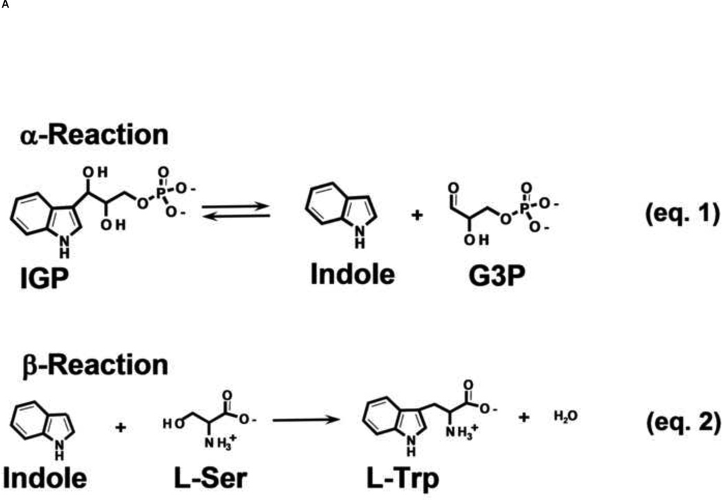
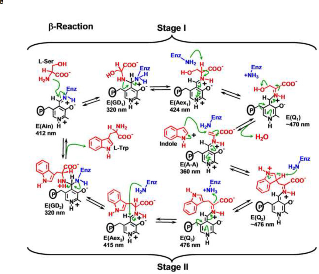
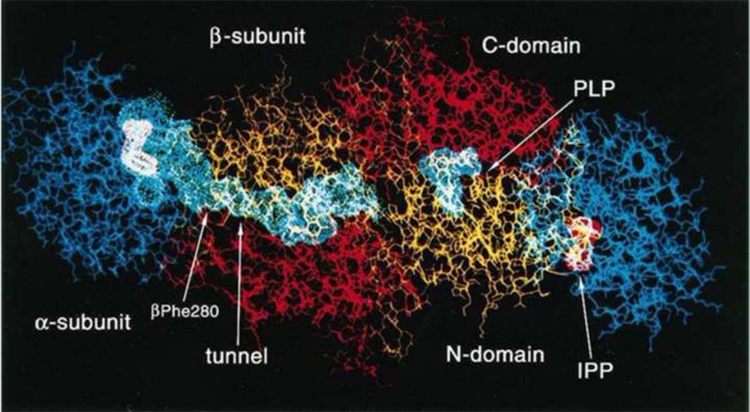
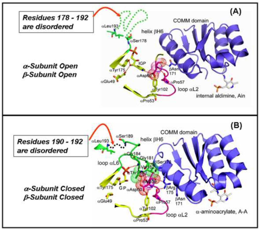
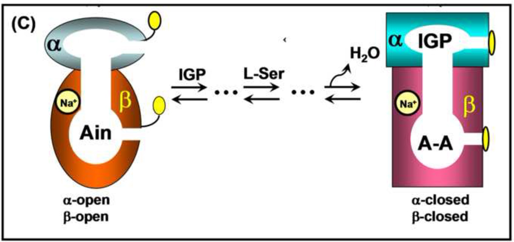
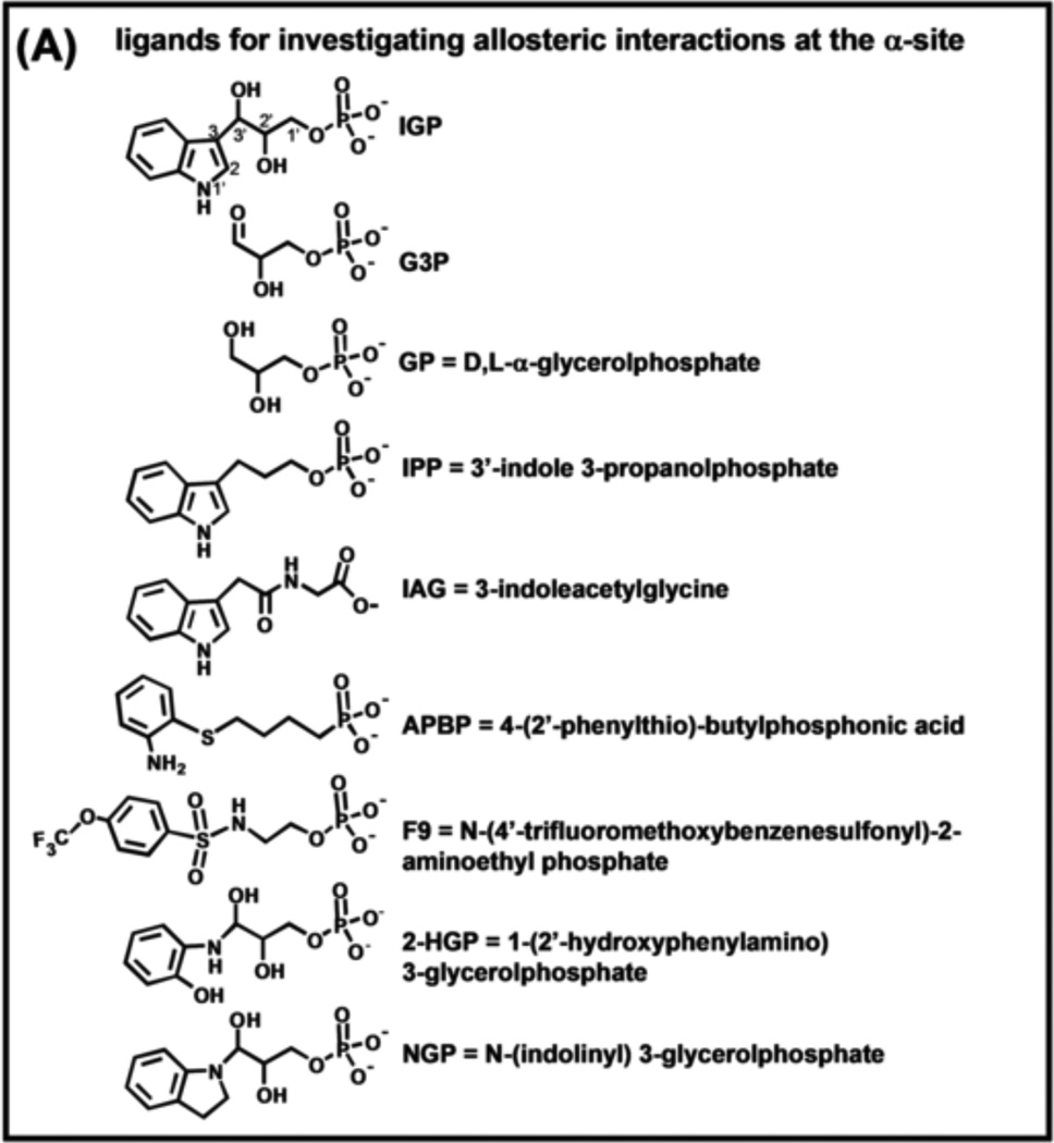
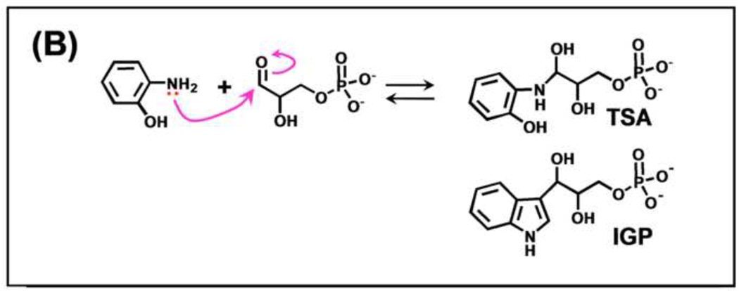
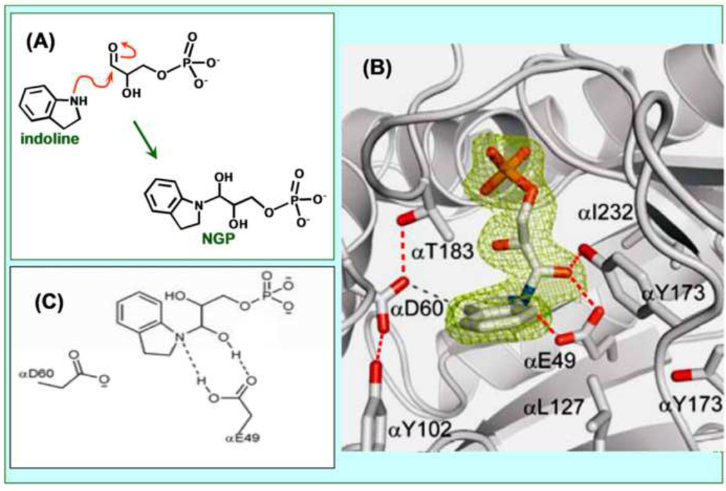
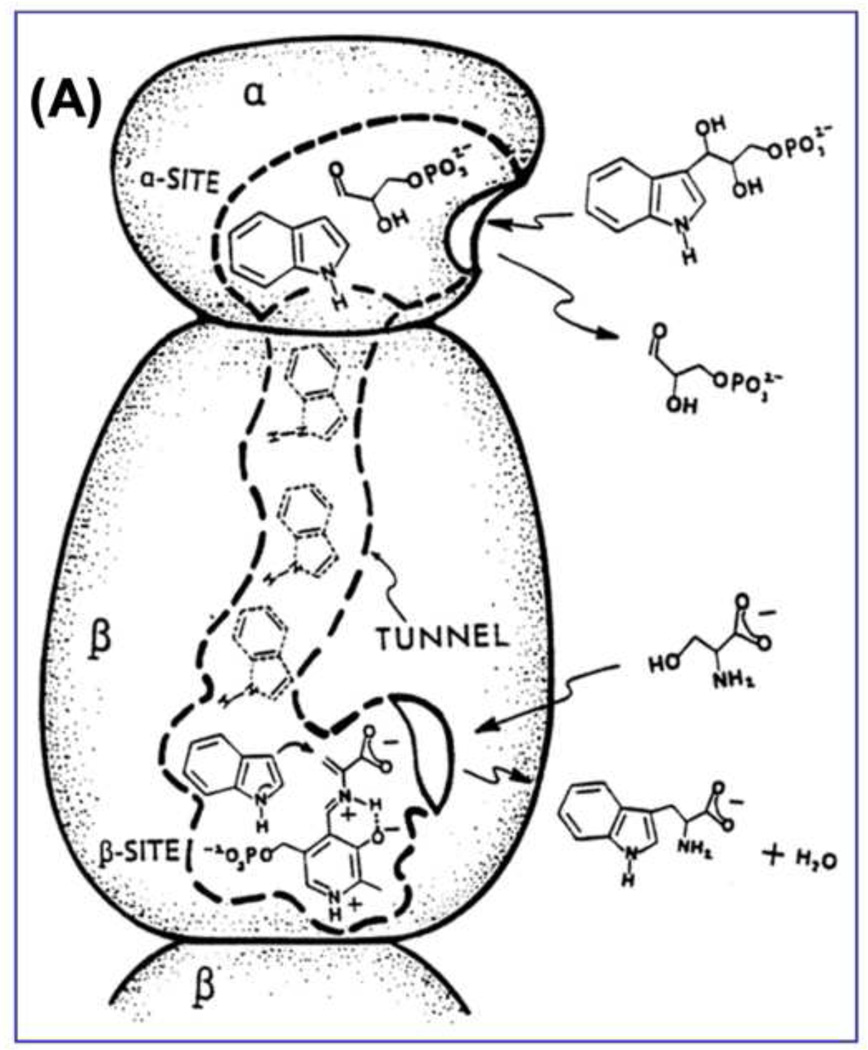
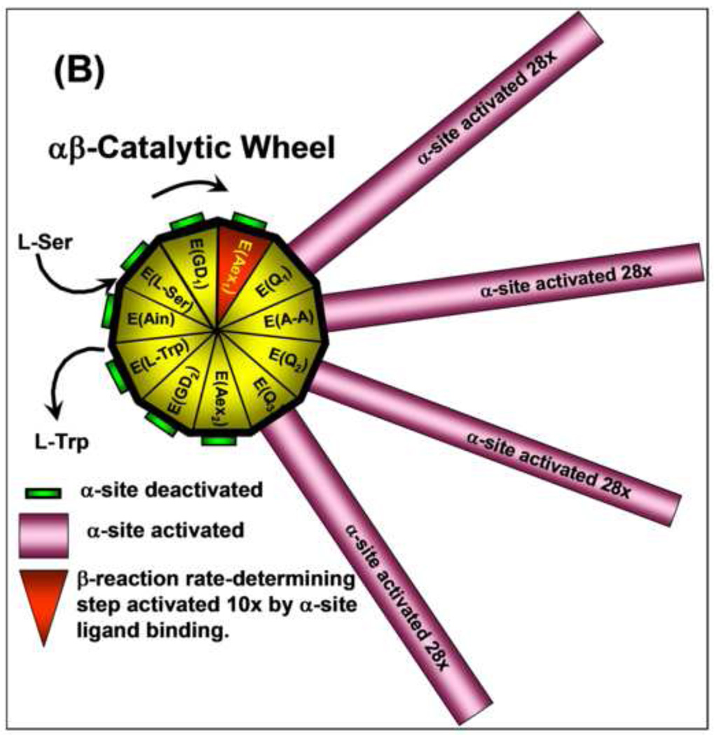
The tryptophan synthase α2β2 bi-enzyme complex catalyzes the last two steps in the synthesis of l-tryptophan (l-Trp). The α-subunit catalyzes cleavage of 3-indole-d-glycerol 3'-phosphate (IGP) to give indole and d-glyceraldehyde 3'-phosphate (G3P). Indole is then transferred (channeled) via an interconnecting 25Å-long tunnel, from the α-subunit to the β-subunit where it reacts with l-Ser in a pyridoxal 5'-phosphate-dependent reaction to give l-Trp and a water molecule. The efficient utilization of IGP and l-Ser by tryptophan synthase to synthesize l-Trp utilizes a system of allosteric interactions that (1) function to switch the α-site on and off at different stages of the β-subunit catalytic cycle, and (2) prevent the escape of the channeled intermediate, indole, from the confines of the α- and β-catalytic sites and the interconnecting tunnel. This review discusses in detail the chemical origins of the allosteric interactions responsible both for switching the α-site on and off, and for triggering the conformational changes between open and closed states which prevent the escape of indole from the bienzyme complex.


Figures

Figure 1
Figure 1
(A) Equations 1 and 2, the α- and β-reactions, respectively, catalyzed by tryptophan synthase [2] (B) Organic chemistry of Stages I and II of the β-reaction. The α-aminoacrylate intermediate, E(A–A), is formed in Stage I. Indole formed in the α-reaction reacts with E(A–A) in Stage II to give L-tryptophan.
Figure 1
Figure 1
(A) Equations 1 and 2, the α- and β-reactions, respectively, catalyzed by tryptophan synthase [2] (B) Organic chemistry of Stages I and II of the β-reaction. The α-aminoacrylate intermediate, E(A–A), is formed in Stage I. Indole formed in the α-reaction reacts with E(A–A) in Stage II to give L-tryptophan.
Figure 2
Figure 2
Structural model for the tryptophan synthase α2β2 bienzyme complex showing important features of the subunits, sites, coenzyme PLP and the interconnecting tunnel. The IGP analogue, indole 3-propanol phosphate (IPP) is bound to the active sites of the α-subunits. Figure modified from Hyde et al. [21].
Figure 3
Figure 3
Structural models depicting components of the conformational transition from the open to the closed αβ-dimeric unit of tryptophan synthase. (A) The αβ-dimeric units of the E(Ain) complex (PDB ID: 1KFK) have the open conformation, whereas, the αβ-dimeric units of the E(A–A) complex have the closed conformation (PDB ID: 2J9X). In (A) and (B), loop αL6 (residues α-176 to α-193) is shown in green. Loop αL2 is shown in magenta. The α-site ligands and residues αE49, αP53, αY102, and αY175 are shown in yellow. van der Waals dot structures are drawn around αA59 and αD60 in (A) and around αA59, αD60, αV182 and αT 183 in (B). Comparison of the structural models in (A) and (B) show that residues α178–192 of loop αL2 are disordered (dashed green line) in the E(Ain) complex (A). In the E(A–A) complex (B), only residues α190–192 of αL2 (dashed green line) are disordered. (C) Cartoon depicting the ligand-mediated interconversion of the open and closed conformations of the αβ-dimeric unit. The combination of IGP binding to the α-site and L-Ser reaction to give E(A–A) at the β-site stabilize the closed conformation of the αβ-dimeric unit. Redrawn from Dunn et al. [27]
Figure 3
Figure 3
Structural models depicting components of the conformational transition from the open to the closed αβ-dimeric unit of tryptophan synthase. (A) The αβ-dimeric units of the E(Ain) complex (PDB ID: 1KFK) have the open conformation, whereas, the αβ-dimeric units of the E(A–A) complex have the closed conformation (PDB ID: 2J9X). In (A) and (B), loop αL6 (residues α-176 to α-193) is shown in green. Loop αL2 is shown in magenta. The α-site ligands and residues αE49, αP53, αY102, and αY175 are shown in yellow. van der Waals dot structures are drawn around αA59 and αD60 in (A) and around αA59, αD60, αV182 and αT 183 in (B). Comparison of the structural models in (A) and (B) show that residues α178–192 of loop αL2 are disordered (dashed green line) in the E(Ain) complex (A). In the E(A–A) complex (B), only residues α190–192 of αL2 (dashed green line) are disordered. (C) Cartoon depicting the ligand-mediated interconversion of the open and closed conformations of the αβ-dimeric unit. The combination of IGP binding to the α-site and L-Ser reaction to give E(A–A) at the β-site stabilize the closed conformation of the αβ-dimeric unit. Redrawn from Dunn et al. [27]
Figure 4
Figure 4
(A) Organic structures and names of some useful α-site ligands. APBP [52], 2-HGP [55] and NGP [27, 47] have been proposed to be transition state analogues for the α-reaction. (B) Reaction of 2-aminophenol with G3P to give the proposed transition state analogue, 2-HGP at the α-site [54]. (C) Stereo structural diagram comparing important features of the α-sites of the complex of 2-HGP (PDB ID: 1TJP) with the complex of IGP (PDB ID: 1QOQ). Color scheme: protein residues of the 2-HGP complex are shown with CPK coloring; 2-HGP, C yellow, N, O, and P in CPK colors. The protein residues of the IGP complex are shown with C orange, N, and O in CPK colors; IGP, C green, N, O, and P in CPK colors. Proposed hydrogen-bonding interactions are show as black dashed lines. Notice that the αD49 carboxylate takes up two orientations in the IGP complex, one with a hydrogen bond between the αD49 carboxylate and the 3-hydroxyl of IGP, the other with the carboxylate rotated by approximately 100° away from IGP.
Figure 4
Figure 4
(A) Organic structures and names of some useful α-site ligands. APBP [52], 2-HGP [55] and NGP [27, 47] have been proposed to be transition state analogues for the α-reaction. (B) Reaction of 2-aminophenol with G3P to give the proposed transition state analogue, 2-HGP at the α-site [54]. (C) Stereo structural diagram comparing important features of the α-sites of the complex of 2-HGP (PDB ID: 1TJP) with the complex of IGP (PDB ID: 1QOQ). Color scheme: protein residues of the 2-HGP complex are shown with CPK coloring; 2-HGP, C yellow, N, O, and P in CPK colors. The protein residues of the IGP complex are shown with C orange, N, and O in CPK colors; IGP, C green, N, O, and P in CPK colors. Proposed hydrogen-bonding interactions are show as black dashed lines. Notice that the αD49 carboxylate takes up two orientations in the IGP complex, one with a hydrogen bond between the αD49 carboxylate and the 3-hydroxyl of IGP, the other with the carboxylate rotated by approximately 100° away from IGP.
Figure 5
Figure 5
(A) Reaction of indoline with G3P at the α-site to form the proposed transition state analogue NGP. (B) Structural model showing NGP fit to the electron density at the α-site (PDB ID: 3CEP). Hydrogen-bonding interactions between active site residues αE49, αD60, αY102 and αT183 and to the amino group of NGP are shown as dashed lines. The α-subunit is shown as a gray cartoon ribbon, active site resides and NGP are shown as sticks with CPK colors. (C) The organic structure of NGP and the hydrogen-bonding interactions to αE49. Redrawn from Dunn et al. [27]. (D) Stereo model depicting the microenvironment of the αE49 carboxyl group in the complex with NGP (PDB ID: 3CEP). Notice that a water molecule (wat291) forms a hydrogen-bonded bridge between the phenolic hydroxyl of αY173 and one oxygen of αE49.
Figure 5
Figure 5
(A) Reaction of indoline with G3P at the α-site to form the proposed transition state analogue NGP. (B) Structural model showing NGP fit to the electron density at the α-site (PDB ID: 3CEP). Hydrogen-bonding interactions between active site residues αE49, αD60, αY102 and αT183 and to the amino group of NGP are shown as dashed lines. The α-subunit is shown as a gray cartoon ribbon, active site resides and NGP are shown as sticks with CPK colors. (C) The organic structure of NGP and the hydrogen-bonding interactions to αE49. Redrawn from Dunn et al. [27]. (D) Stereo model depicting the microenvironment of the αE49 carboxyl group in the complex with NGP (PDB ID: 3CEP). Notice that a water molecule (wat291) forms a hydrogen-bonded bridge between the phenolic hydroxyl of αY173 and one oxygen of αE49.
Figure 6
Figure 6
The step-wise and concerted mechanisms proposed for the α-reaction are presented in (A) and (B) respectively. In both mechanisms, αE49 plays the role of acid-base catalyst for the proton transfers required for scission of the IGP C-C bond, while αD60 plays a charge-charge role in stabilizing charge development on the indole ring N.
Figure 7
Figure 7
Panels (A) – (D) present stereo diagrams comparing structural models for the β-active site and the COMM domain (residues β102-β189) of the open and closed β- subunit conformations. (A) Open conformation of the E(Aex1) complex (PDB ID: 2CLL). The β-subunit COMM domain of E(Aex1) is shown as a cartoon ribbon (green), active site residues and the L-Ser moiety (Aex1) bound to PLP are shown as sticks with CPK coloring. In this structure [45] the β-hydroxyl of the Aex1 moiety (black arrow) is hydrogen-bonded (black dashed line) to the carboxylate of βD305. This interaction stabilizes the inactive (open) conformation of E(Aex1) by making removal of the proton chemically more difficult and by preventing bonding interaction(s) with the acid-base catalytic groups at the site, βK87 and βE109. The models in (B) compare the open structure of E(Aex1) from (A) with the closed structure of the α-aminoacrylate intermediate, E(A–A) (PDB ID: 2J9X). The closed structure of the E(A–A) complex is superimposed on the E(Aex1) structure. The COMM domain of E(A–A) is shown as a magenta cartoon ribbon with the active site residues and PLP-bound α-aminoacrylate depicted as stick structures with carbons yellow, and with O, N, and P in CPK colors. Water molecule, wat88, is shown as a red ball, located near the β-carbon of the α-aminoacrylate moiety. Dashed black lines indicate hydrogen bonds. For clarity, the structure of the closed conformation of the E(A–A) complex from (B) is shown in (C). The motion of the COMM domain in the transition from the open to the closed conformation brings βR141 and βD305 together to form a salt bridge along with new hydrogen bonding interactions to βS297 and βS299 [46]. Hydrogen bonds and the distance between the water molecule and the β-carbon of the α-aminoacrylate moiety, (A–A), are indicated by black dashed lines. The βR141-βD305 salt bridge provides a physical barrier which prevents the transfer of ligands between solution and the β- site. This closed conformation also positions βE109 and βK87 for the proton transfers required in the catalytic steps which take place in Stage II of the β- reaction [27, 46, 47, 49]. (D) Stereo structural model showing the closed β-site of the βK87T variant with the L-Trp external aldimine, E(Aex2), bound to the β-site. Notice that the carboxylate of βE109 is hydrogen-bonded to the indole ring N of Aex2 (black dashed line) (PDB ID: 2TYS).
Figure 7
Figure 7
Panels (A) – (D) present stereo diagrams comparing structural models for the β-active site and the COMM domain (residues β102-β189) of the open and closed β- subunit conformations. (A) Open conformation of the E(Aex1) complex (PDB ID: 2CLL). The β-subunit COMM domain of E(Aex1) is shown as a cartoon ribbon (green), active site residues and the L-Ser moiety (Aex1) bound to PLP are shown as sticks with CPK coloring. In this structure [45] the β-hydroxyl of the Aex1 moiety (black arrow) is hydrogen-bonded (black dashed line) to the carboxylate of βD305. This interaction stabilizes the inactive (open) conformation of E(Aex1) by making removal of the proton chemically more difficult and by preventing bonding interaction(s) with the acid-base catalytic groups at the site, βK87 and βE109. The models in (B) compare the open structure of E(Aex1) from (A) with the closed structure of the α-aminoacrylate intermediate, E(A–A) (PDB ID: 2J9X). The closed structure of the E(A–A) complex is superimposed on the E(Aex1) structure. The COMM domain of E(A–A) is shown as a magenta cartoon ribbon with the active site residues and PLP-bound α-aminoacrylate depicted as stick structures with carbons yellow, and with O, N, and P in CPK colors. Water molecule, wat88, is shown as a red ball, located near the β-carbon of the α-aminoacrylate moiety. Dashed black lines indicate hydrogen bonds. For clarity, the structure of the closed conformation of the E(A–A) complex from (B) is shown in (C). The motion of the COMM domain in the transition from the open to the closed conformation brings βR141 and βD305 together to form a salt bridge along with new hydrogen bonding interactions to βS297 and βS299 [46]. Hydrogen bonds and the distance between the water molecule and the β-carbon of the α-aminoacrylate moiety, (A–A), are indicated by black dashed lines. The βR141-βD305 salt bridge provides a physical barrier which prevents the transfer of ligands between solution and the β- site. This closed conformation also positions βE109 and βK87 for the proton transfers required in the catalytic steps which take place in Stage II of the β- reaction [27, 46, 47, 49]. (D) Stereo structural model showing the closed β-site of the βK87T variant with the L-Trp external aldimine, E(Aex2), bound to the β-site. Notice that the carboxylate of βE109 is hydrogen-bonded to the indole ring N of Aex2 (black dashed line) (PDB ID: 2TYS).
Figure 8
Figure 8
(A) Cartoon for the αβ-dimeric unit of tryptophan synthase depicting the binding and dissociation of ligands to the α- and β-sites and the transfer of intermediate indole from the α-site to the β-site (redrawn from [62]). (B) Catalytic wheel summarizing the interplay of ligand binding and reaction with the allosteric signaling between the α- and β-sites during the catalytic cycles of the α- and β-reactions. The gold and red triangular segments of the wheel hub represent the different known chemical states of the β-catalytic cycle (see Figure 1). The green or magenta bars extending out from each triangular segment have lengths proportional to the relative activity of the α-site for each chemical state of the β-site. As the E(Ain) species is converted by reaction with L-Ser to E(GD1) and E(Aex1) the α-site remains in a catalytically inactive state. When E(Aex1) is converted to E(A–A), via E(Q1), the α-site is activated at least 28-fold [61], and remains activated until E(Q3) is converted to E(Aex2) when the α-site is switched off again [63]. As the β-reaction continues from E(Aex2) to the complex of E(Ain) with L-Trp, the α-site remains deactivated. The triangular segment designated as E(Aex1) is colored red to signify activation of the conversion of this intermediate to E(A–A) by IGP binding to the α-site [27]. Modulation of the switching of the α- and β-sites between open states of low activity and closed states of high activity by ligand binding and reaction provides a mechanism for both synchronizing the α- and β-reactions and preventing the escape of indole from the confines of the α- and β-sites and the interconnecting tunnel. This conformational switching makes possible the efficient channeling of indole during the αβ-catalytic cycle of tryptophan synthase. Figure redrawn from [27].
Figure 8
Figure 8
(A) Cartoon for the αβ-dimeric unit of tryptophan synthase depicting the binding and dissociation of ligands to the α- and β-sites and the transfer of intermediate indole from the α-site to the β-site (redrawn from [62]). (B) Catalytic wheel summarizing the interplay of ligand binding and reaction with the allosteric signaling between the α- and β-sites during the catalytic cycles of the α- and β-reactions. The gold and red triangular segments of the wheel hub represent the different known chemical states of the β-catalytic cycle (see Figure 1). The green or magenta bars extending out from each triangular segment have lengths proportional to the relative activity of the α-site for each chemical state of the β-site. As the E(Ain) species is converted by reaction with L-Ser to E(GD1) and E(Aex1) the α-site remains in a catalytically inactive state. When E(Aex1) is converted to E(A–A), via E(Q1), the α-site is activated at least 28-fold [61], and remains activated until E(Q3) is converted to E(Aex2) when the α-site is switched off again [63]. As the β-reaction continues from E(Aex2) to the complex of E(Ain) with L-Trp, the α-site remains deactivated. The triangular segment designated as E(Aex1) is colored red to signify activation of the conversion of this intermediate to E(A–A) by IGP binding to the α-site [27]. Modulation of the switching of the α- and β-sites between open states of low activity and closed states of high activity by ligand binding and reaction provides a mechanism for both synchronizing the α- and β-reactions and preventing the escape of indole from the confines of the α- and β-sites and the interconnecting tunnel. This conformational switching makes possible the efficient channeling of indole during the αβ-catalytic cycle of tryptophan synthase. Figure redrawn from [27].

References

    1. Bertrand K, Korn L, Lee F, Platt T, Squires CL, Squires C, Yanofsky C. New features of the regulation of the tryptophan operon. Science. 1975;189:22–26. - PubMed
    1. Yanofsky C, Crawford IP. In: Tryptophan synthase. 3rd ed. Boyer PD, editor. New York: The Enzymes, Academic Press; 1972. pp. 1–31.
    1. Miles EW. Tryptophan synthase: structure, function, and subunit interaction. Adv. Enzymol. Relat Areas Mol. Biol. 1979;49:127–186. - PubMed
    1. Umbreit W, Wood W, Gunsalus I. The activity of pyridoxal phosphate in tryptophan formation by cell-free enzyme preparations. J. Biol. Chem. 1946;165:731–732. - PubMed
    1. Yanofsky C. Tryptophan synthetase from Neutrospora. In: Colowick SP, Kaplan NO, editors. Methods of Enzymol. vol. II. N. Y.: Academic Press, Inc.; 1995. pp. 233–238.

Publication types

MeSH terms

Substances

LinkOut - more resources