

Age-related changes in osseous anatomy, alignment, and range of motion of the cervical spine. Part I: Radiographic data from over 1,200 asymptomatic subjects
- PMID: 22310883
- PMCID: PMC3535253
- DOI: 10.1007/s00586-012-2167-5
Age-related changes in osseous anatomy, alignment, and range of motion of the cervical spine. Part I: Radiographic data from over 1,200 asymptomatic subjects
Abstract
Purpose: This study aimed to establish radiographic standard values for cervical spine morphometry, alignment, and range of motion (ROM) in both male and female in each decade of life between the 3rd and 8th and to elucidate these age-related changes.
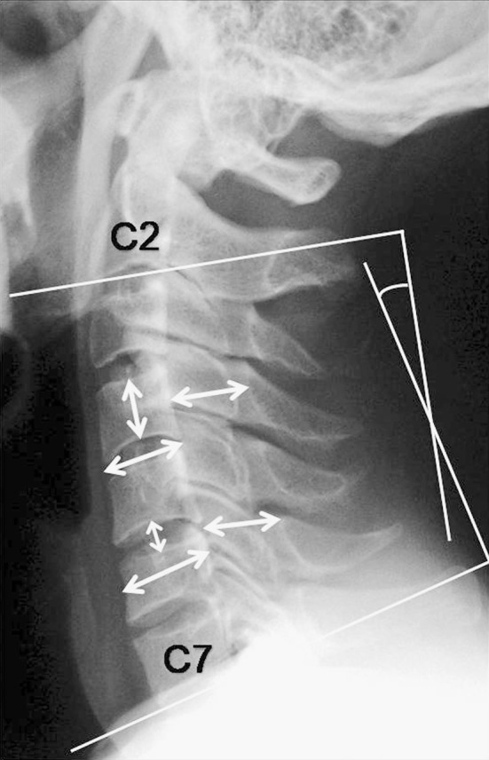
Methods: A total of 1,230 asymptomatic volunteers underwent anteroposterior (AP), lateral, flexion, and extension radiography of the cervical spine. There were at least 100 men and 100 women in each decade of life between the 3rd and 8th. AP diameter of the spinal canal, vertebral body, and disc were measured at each level from the 2nd to 7th cervical vertebra (C2-C7). C2-C7 sagittal alignment and ROM during flexion and extension were calculated using a computer digitizer.
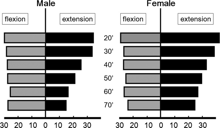
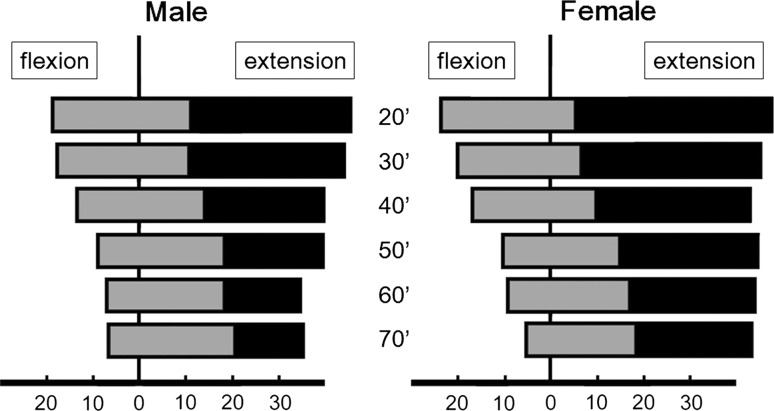
Results: The AP diameter of the spinal canal was 15.8 ± 1.5 [mean ± standard deviation (SD)] mm at the mid-C5 level, and 15.5 ± 2.0 mm at the C5/6 disc level. The disc height was 5.8 ± 1.3 mm at the C5/6 level, which was the minimum height, and the maximum height was at the C6/7 level. Both the AP diameter of the spinal canal and disc height decreased gradually with increasing age. The C2-C7 sagittal alignment and total ROM were 13.9 ± 12.3° in lordosis and 55.3 ± 16.0°, respectively. The C2-C7 lordotic angle was 8.0 ± 11.8° in the 3rd decade and increased to 19.7 ± 11.3 in the 8th decade, whereas the C2-C7 ROM was 67.7 ± 17.0° in the 3rd decade and decreased to 45.0 ± 12.5 in the 8th decade. The extension ROM decreased more than the flexion ROM, and lordotic alignment progressed with increasing age. There was a significant difference in C2-C7 alignment and ROM between men and women.
Conclusions: The standard values and age-related changes in cervical anatomy, alignment, and ROM for males and females in each decade between the 3rd and 8th were established. Cervical lordosis in the neutral position develops with aging, while extension ROM decreases gradually. These data will be useful as normal values for the sake of comparison in clinical practice.
Figures
References
-
- Ferlic D. The range of motion of the “normal” cervical spine. Bull Johns Hopkins Hosp. 1962;110:59–65. - PubMed
-
- Fon GT, Pitt MJ, Thies AC., Jr Thoracic kyphosis: range in normal subjects. AJR Am J Roentgenol. 1980;134(5):979–983. - PubMed
-
- Hinck VC, Hopkins CE, Savara BS. Sagittal diameter of the cervical spinal canal in children. Radiology. 1962;79:97–108. - PubMed
Publication types
MeSH terms
LinkOut - more resources
Full Text Sources
Medical
Research Materials
Miscellaneous
