. 2012 Mar 23;287(13):10565-10578.
doi: 10.1074/jbc.M111.294645. Epub 2012 Feb 6.

Phospholipase C-related but catalytically inactive protein (PRIP) modulates synaptosomal-associated protein 25 (SNAP-25) phosphorylation and exocytosis

Affiliations

Phospholipase C-related but catalytically inactive protein (PRIP) modulates synaptosomal-associated protein 25 (SNAP-25) phosphorylation and exocytosis

Jing Gao et al. J Biol Chem. .

Abstract

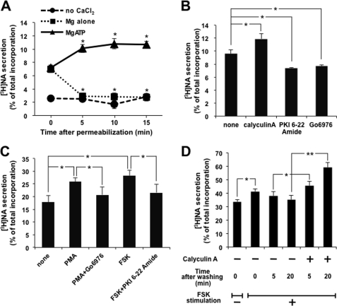
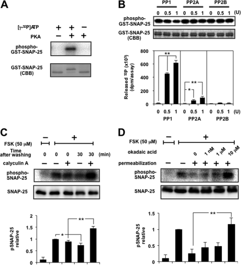
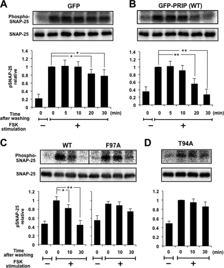
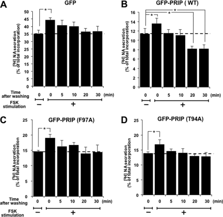
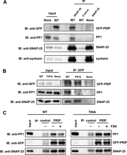
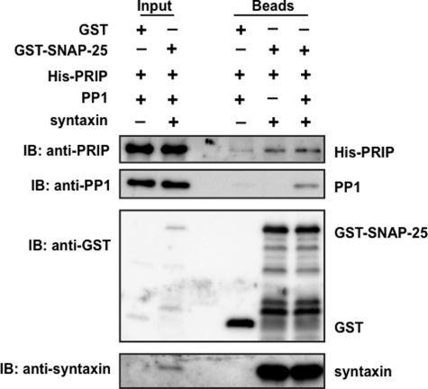
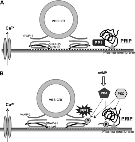
Exocytosis is one of the most fundamental cellular events. The basic mechanism of the final step, membrane fusion, is mediated by the formation of the SNARE complex, which is modulated by the phosphorylation of proteins controlled by the concerted actions of protein kinases and phosphatases. We have previously shown that a protein phosphatase-1 (PP1) anchoring protein, phospholipase C-related but catalytically inactive protein (PRIP), has an inhibitory role in regulated exocytosis. The current study investigated the involvement of PRIP in the phospho-dependent modulation of exocytosis. Dephosphorylation of synaptosome-associated protein of 25 kDa (SNAP-25) was mainly catalyzed by PP1, and the process was modulated by wild-type PRIP but not by the mutant (F97A) lacking PP1 binding ability in in vitro studies. We then examined the role of PRIP in phospho-dependent regulation of exocytosis in cell-based studies using pheochromocytoma cell line PC12 cells, which secrete noradrenalin. Exogenous expression of PRIP accelerated the dephosphorylation process of phosphorylated SNAP-25 after forskolin or phorbol ester treatment of the cells. The phospho-states of SNAP-25 were correlated with noradrenalin secretion, which was enhanced by forskolin or phorbol ester treatment and modulated by PRIP expression in PC12 cells. Both SNAP-25 and PP1 were co-precipitated in anti-PRIP immunocomplex isolated from PC12 cells expressing PRIP. Collectively, together with our previous observation regarding the roles of PRIP in PP1 regulation, these results suggest that PRIP is involved in the regulation of the phospho-states of SNAP-25 by modulating the activity of PP1, thus regulating exocytosis.


Figures

FIGURE 1.
FIGURE 1.
Protein phosphorylation is involved in the regulation of exocytosis. A, PC12 cells labeled with [3H]NA were permeabilized with 10 μm digitonin and then maintained at room temperature for the period indicated in the presence or absence of 2 mm MgATP. [3H]NA secretion was triggered by adding 2 mm CaCl2 to give a free Ca2+ concentration of 1 μm for 2 min. NA release is indicated as the percentage of released [3H]NA relative to the total [3H]NA incorporated into the cells. B, after digitonin permeabilization, [3H]NA-labeled PC12 cells were incubated at room temperature for 5 min in the presence of 2 mm MgATP, along with either calyculin A (100 nm), PKI 6-22 amide (100 nm), or Go6976 (1 μm). [3H]NA secretion by 1 μm Ca2+ for 2 min was assayed. C, PC12 cells labeled with [3H]NA were treated with PMA (1 μm) or forskolin (FSK) (50 μm) with or without each inhibitor for 30 or 5 min, respectively, followed by the assay of [3H]NA secretion by high K+ solution (70 mm KCl) for 2 min. D, PC12 cells labeled with [3H]NA were treated with 50 μm FSK for 5 min. After removal of the stimulus, cells were left for 5 or 20 min in the presence or absence of 100 nm calyculin A, followed by the assay of [3H]NA secretion by 10 μm ionomycin for 5 min. All of the results are the means ± S.E. (error bars) of five independent experiments. Student's t tests were used, and significance is represented by * or ** for p < 0.05 or p < 0.01, respectively. The same statistical methods were applied to the following results.
FIGURE 2.
FIGURE 2.
Dephosphorylation of SNAP-25 was mainly catalyzed by PP1. A, GST-tagged SNAP-25 was phosphorylated with [γ-32P]ATP using the catalytic subunit of PKA. The mixture was separated by SDS-PAGE, followed by CBB staining and autoradiography. Note that phosphorylated SNAP-25 shifted up. B, GST-tagged SNAP-25 immobilized on glutathione beads was phosphorylated as described above, followed by dephosphorylation by PP1, PP2A, or PP2B in an appropriate buffer solution. The radioactivity of released 32P was counted using a liquid scintillation counter, and beads were analyzed by SDS-PAGE for CBB staining and autoradiography. The top and bottom panels show a typical autoradiogram and CBB staining of GST-SNAP-25 after treatment, respectively. The graph shows a summary of the released radioactivity of five separate experiments. Doublet bands of SNAP-25 seen by CBB staining showed little difference after treatment with PP, indicating that liberation of radioactivity is more sensitive, and the portion of SNAP-25 dephosphorylated appears to be small. Each enzyme was supplied by the respective manufacturer, designating the activity as a unit, but the assay condition (buffer composition, time, temperature, etc.) was different from that used in the present study, so we re-evaluated each activity using 100 μm p-nitrophenyl phosphate as a substrate under exactly the same assay conditions as described here. One unit of PP1, PP2A, and PP2B by the manufacturer was about 0.3, 0.7, and 0.2 unit, respectively. Re-evaluated activity of each enzyme was used here. C, PC12 cells labeled with [32P]orthophosphate were stimulated with 50 μm FSK for 5 min. After removal of the stimulus, cells were left in the presence or absence of 100 nm calyculin A for 30 min. Cellular extracts were subjected to immunoprecipitation by anti-SNAP-25 antibody overnight at 4 °C, and the immunocomplexes were examined by SDS-PAGE followed by autoradiography and Western blotting using anti-SNAP-25 antibody. The panels show a typical autoradiogram and immunoblot, and the graph shows the summary of five separate experiments. Doublet bands of SNAP-25 depending on the phospho-state seen by CBB staining in vitro (see A and B) were not observed in that immunoprecipitated by anti-SNAP-25 antibody from living cells, for unknown reasons. D, PC12 cells labeled with [32P]orthophosphate were incubated with 50 μm FSK for 5 min, followed by digitonin permeabilization and subsequent incubation with okadaic acid at the concentrations indicated for another 10 min. The cell lysates were subjected to immunoprecipitation as described above. The panels show a typical autoradiogram and immunoblot, and the results are expressed as the mean ± S.E. (error bars) of five separate experiments.
FIGURE 3.
FIGURE 3.
Dephosphorylation of SNAP-25 is modulated by PRIP-1 in vitro. GST-tagged SNAP-25 immobilized on glutathione beads was phosphorylated as described and then dephosphorylated by 1 unit of PP1. A, recombinant PRIP-1 (WT) or the mutant F97A (1 nmol) was included in the reaction mixture. The panels show an autoradiogram and CBB staining of GST-SNAP-25 after treatment with PP1 alone or along with PRIP-1. Results are expressed as the mean ± S.E. of five separate experiments. CBB staining of SNAP-25 yielded double bands; upper and lower bands represent phosphorylated and unphosphorylated, respectively (see Fig. 2A). B, PRIP-1 was phosphorylated in advance by non-radioactive ATP plus the catalytic subunit of PKA (pPRIP), followed by a dephosphorylation assay of SNAP-25. In this experiment, radioactivity to phosphorylate SNAP-25 was reduced. Results are expressed as the mean ± S.E. (error bars) of four separate experiments.
FIGURE 4.
FIGURE 4.
Dephosphorylation of SNAP-25 was modulated in PC12 cells expressing PRIP-1. PC12 cells expressing GFP (A), GFP-PRIP-1 (WT) (B), GFP-RPIP-1 (F97A) (C), or GFP-PRIP-1 (T94A) (D) were labeled with [32P]orthophosphate, followed by stimulation with 50 μm FSK for 5 min. After removal of the stimulus, cells were left for the time period indicated. Cell extracts were subjected to immunoprecipitation by anti-SNAP-25 antibody overnight at 4 °C, and the precipitates were examined by SDS-PAGE for autoradiography and Western blotting using anti-SNAP-25 antibody. The panels show a typical autoradiogram and immunoblot. Results are expressed as the mean ± S.E. (error bars) of five (A–C) or four (D) separate experiments.
FIGURE 5.
FIGURE 5.
[3H]NA secretion was modulated by PRIP-1. PC12 cells expressing GFP (A), GFP-PRIP-1 (WT) (B), GFP-RPIP-1 (F97A) (C), or GFP-PRIP-1 (T94A) (D) were labeled with [3H]NA, followed by stimulation with 50 μm FSK for 5 min. After removal of the stimulus, cells were left at room temperature for 5, 10, 20, or 30 min, followed by [3H]NA secretion assay with high K+ solution for 5 min. All the data are the means ± S.E. (error bars) of five (A–C) or three (D) experiments.
FIGURE 6.
FIGURE 6.
Interaction of PRIP-1, SNAP-25, and PP1 in PC12 cells. A, PC12 cells expressing GFP alone (None) or GFP-PRIP-1 (WT) were lysed, and SNAP-25 was immunoprecipitated (IP) by anti-SNAP25 antibody. Immunoprecipitate was separated by SDS-PAGE and analyzed by Western blotting (IB) using anti-GFP, anti-PP1, anti-SNAP25, and anti-syntaxin antibodies. Control, immunoprecipitation by control IgG instead of anti-SNAP-25 antibody. Similar results were seen in three other independent experiments. 0.1 or 30% of the total amounts of cell lysates or immunoprecipitates, respectively, was applied to SDS-PAGE, the values of which were taken into account for the calculation (see “Results”). B, PC12 cells expressing GFP (None), GFP-PRIP-1 (WT), or GFP-PRIP-1 (F97A) were lysed and subjected to immunoprecipitation with anti-GFP antibody using the Pierce Crosslink Immunoprecipitation Kit. The eluted samples were separated by SDS-PAGE and analyzed by Western blotting using anti-GFP, anti-SNAP25, and anti-PP1 antibody. Similar results were seen in three other independent experiments. C, PC12 cells expressing GFP-PRIP-1 (WT) or GFP-PRIP-1 (T94A) were first stimulated to phosphorylate PRIP-1 itself with FSK (50 μm) for 5 min, followed by immunoprecipitation by anti-PRIP-1 antibody (PRIP). The immunoprecipitates were immunoblotted by anti-PP1 and anti-SNAP-25 antibodies. Control, immunoprecipitation by control IgG instead of anti-PRIP-1. The band seen in the blot by anti-SNAP-25 in the control would be a light chain of IgG because a band with a similar density was observed even in the absence of cell lysates. Similar results were seen in two other independent experiments.
FIGURE 7.
FIGURE 7.
In vitro complex formation of SNAP-25 with PRIP, PP1 and syntaxin. An equal amount of each molecule was assayed for the complex formation. GST alone or GST-SNAP-25 along with His-PRIP-1, PP1, and syntaxin at the same amount (10 pmol) was incubated at 4 °C for 2 h, followed by the addition of excess capacity of glutathione beads, precipitation by centrifugation, separation by SDS-PAGE, and immunoblotting (IB) with each antibody. A typical blot is shown, and two other blots yielded similar results. Bands seen between GST-SNAP-25 and GST by immunoblotting with anti-GST antibody would be degradative products of GST-SNAP-25. 0.1 or 30% of the recombinant molecules used or beads after experiments, respectively, were applied to SDS-PAGE, the values of which were taken into account for the calculation (see “Results”).
FIGURE 8.
FIGURE 8.
Schematic representation of the role of PRIP in phospho-dependent regulation of SNAP-25. A, under basal conditions, PRIP facilitates the targeting of PP1 to the site where SNAP-25 exists. PP1 bound by PRIP is inactive. B, PKA or PKC triggers the phosphorylation of SNAP-25 and PRIP, thus modulating exocytosis. Phosphorylated PRIP releases PP1 to be activated for dephosphorylation to cease phospho-modulation by SNAP-25. The triangle indicates the devices available for PRIP to approach the site where exocytosis occurs (see “Discussion”).

References

    1. Banerjee A., Barry V. A., DasGupta B. R., Martin T. F. (1996) N-Ethylmaleimide-sensitive factor acts at a prefusion ATP-dependent step in Ca2+-activated exocytosis. J. Biol. Chem. 271, 20223–20226 - PubMed
    1. Sutton R. B., Fasshauer D., Jahn R., Brunger A. T. (1998) Crystal structure of a SNARE complex involved in synaptic exocytosis at 2.4 Å resolution. Nature 395, 347–353 - PubMed
    1. Weber T., Zemelman B. V., McNew J. A., Westermann B., Gmachl M., Parlati F., Söllner T. H., Rothman J. E. (1998) SNAREpins. Minimal machinery for membrane fusion. Cell 92, 759–772 - PubMed
    1. Jahn R., Lang T., Südhof T. C. (2003) Membrane fusion. Cell 112, 519–533 - PubMed
    1. Söllner T. H. (2003) Regulated exocytosis and SNARE function (Review). Mol. Membr. Biol. 20, 209–220 - PubMed

Publication types

MeSH terms

LinkOut - more resources