. 2012 Feb 6;196(3):315-26.
doi: 10.1083/jcb.201107058.

Inhibition of pyrimidine synthesis reverses viral virulence factor-mediated block of mRNA nuclear export

Affiliations

Inhibition of pyrimidine synthesis reverses viral virulence factor-mediated block of mRNA nuclear export

Liang Zhang et al. J Cell Biol. .

Abstract

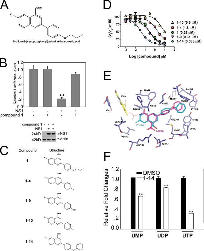
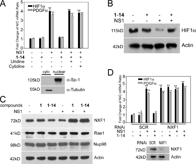
The NS1 protein of influenza virus is a major virulence factor essential for virus replication, as it redirects the host cell to promote viral protein expression. NS1 inhibits cellular messenger ribonucleic acid (mRNA) processing and export, down-regulating host gene expression and enhancing viral gene expression. We report in this paper the identification of a nontoxic quinoline carboxylic acid that reverts the inhibition of mRNA nuclear export by NS1, in the absence or presence of the virus. This quinoline carboxylic acid directly inhibited dihydroorotate dehydrogenase (DHODH), a host enzyme required for de novo pyrimidine biosynthesis, and partially reduced pyrimidine levels. This effect induced NXF1 expression, which promoted mRNA nuclear export in the presence of NS1. The release of NS1-mediated mRNA export block by DHODH inhibition also occurred in the presence of vesicular stomatitis virus M (matrix) protein, another viral inhibitor of mRNA export. This reversal of mRNA export block allowed expression of antiviral factors. Thus, pyrimidines play a necessary role in the inhibition of mRNA nuclear export by virulence factors.


Figures

Figure 1.
Figure 1.
Quinoline carboxylic acid targets DHODH and reverts host gene expression block induced by NS1. (A) Structure of 1, a quinoline carboxylic acid. (B) Luciferase reporter gene assay was performed in 293T cells transfected with a plasmid encoding luciferase alone or cotransfected with a plasmid encoding NS1, in the absence or presence of 5 µM 1 as indicated. NS1 and β-actin levels were measured by immunoblot analysis. (C) Analogues of 1. (D) Dose–response curves for inhibition of human DHODH. Inhibitor concentration was varied in a threefold dilution series (0.01–100 µM), and the percentage of inhibition relative to no inhibitor control was determined (vi/vo × 100). Data collected in triplicate were fitted to the log[I] versus response (three parameters) equation in Prism to determine the IC50. Values given in parenthesis are the fitted IC50 values. A77 1726, the active metabolite of leflunomide, was used as a control and gave an IC50 = 0.25 (0.13–0.47) µM. (E) Model of 1-14 bound to human DHODH generated by DOCK. 1-14 (turquoise) was docked into the human DHODH structure (purple). ABBA, the original ligand cocrystallized with DHODH in 2B0M, is displayed in magenta, flavin mononucleotide (FMN) is displayed in yellow, orotate (oro) is displayed in purple, nitrogens are in blue, and oxygens are in red. Active site residues are labeled. ABBA is a brequinar analogue. (F) Relative abundance of uridine nucleotides determined by liquid chromatography MS/MS in HBECs in the absence or presence of 1-14. Intracellular UTP, UDP, and UMP levels were quantitated using MRM. Results represent mean values ± SD; **, P < 0.01.
Figure 2.
Figure 2.
Quinoline carboxylic acid inhibits virus replication via DHODH. (A) HBECs were noninfected or infected with A/WSN/1933 at an MOI of 1. Cells were pretreated for 2 h with 1-14 and/or posttreated with 1-14, added at the indicated time points, for 24 h. Cell extracts were subjected to immunoblot analysis using antibodies against various influenza virus proteins. (B) HBECs were infected with A/WSN/1933 at an MOI of 0.01, and 1-14, at various concentrations, was added 1 h after infection. Virus replication was measured by plaque assays. (C) MDCK cells were infected with A/WSN/1933 at an MOI of 0.001 for 48 h and incubated with DMSO, 1, 1-10, and 1-14 in the absence (control) or presence of 500 µM uridine (U), cytidine (C), adenosine (A), or guanosine (G). Virus titers were measured by plaque assays. (D) MDCK cells were infected with A/WSN/1933 at an MOI of 0.001 for 48 h and incubated with DMSO or 1-14 in the absence or presence of 50 or 500 µM orotic acid. Virus titers were measured by plaque assays. Results represent mean values ± SD; **, P < 0.01. p.i., postinfection; pfu, plaque-forming unit.
Figure 3.
Figure 3.
Quinoline carboxylic acid releases mRNA export block induced by influenza virus. (A) HBECs were infected with A/WSN/1933 or A/WSN/1933-ΔNS1 at an MOI of 1 for 30 h in the absence or presence of 1 µM 1-14. Cells were subjected to oligo d(T) in situ hybridization and immunofluorescence using antibodies against all influenza virus proteins. Cells were visualized in an inverted microscope (Axiovert 200M). DAPI stained the nucleus. Bar, 20 µM. (B) Quantification of cells that presented nuclear retention of poly(A)+ RNA in A. The percentage of cells presenting nuclear retention of poly(A)+ RNA was calculated with respect to cells that showed normal distribution of poly(A)+ RNA—in the nucleus and in the cytoplasm. 500 cells were counted for each condition. Results represent mean values ± SD; **, P < 0.01.
Figure 4.
Figure 4.
Pyrimidine biosynthesis regulates NS1-targeted host mRNA nuclear export. (A) A luciferase gene expression assay was performed in 293T cells by cotransfection of reporter plasmids and plasmids encoding NS1. Cells were treated with compounds 1 (5 µM) or 1-14 (1 µM) in the presence or absence of uridine or cytidine, as indicated, and luciferase activity was measured. Cell lysates as in A were subjected to immunoblot analysis with antibodies against the indicated proteins. (B) HeLa cells transfected with plasmid alone or plasmid expressing NS1 were incubated with DMSO, 1 µM compound 1-14 alone, or compound 1-14 in the presence of 500 µM uridine. Cells were then subjected to oligo d(T) in situ hybridization and immunofluorescence with anti-NS1 antibodies. Bar, 15 µM. (C) Quantification of cells that presented nuclear retention of poly(A)+ RNA in B. The percentage of cells presenting nuclear retention of poly(A)+ RNA was calculated with respect to cells that showed normal distribution of poly(A)+ RNA—in the nucleus and in the cytoplasm. 500 cells were counted for each condition. Results represent mean values ± SD; **, P < 0.01.
Figure 5.
Figure 5.
DHODH inhibitor induces nuclear export and expression of mRNAs encoding antiviral factors via increasing NXF1 levels. (A) 293T cells transfected with a plasmid encoding NS1 were untreated or treated with 1-14. RNA was isolated from nuclear and cytoplasmic (cyto) fractions, and the indicated mRNA species were quantified by real-time RT-PCR. Controls for fraction purity are shown by immunoblot analysis, as indicated. (B) 293T cells were transfected with a control plasmid or a plasmid encoding NS1 and untreated or treated with 1-14. Total cell extracts were subjected to immunoblot analysis with antibodies against the indicated proteins. (C) 293T cells were transfected with a control plasmid or a plasmid encoding NS1 and untreated or treated with 1 or 1-14. Total cell extracts were subjected to immunoblot analysis with antibodies against the indicated proteins. (D) 293T cells were cotransfected with a control plasmid or a plasmid encoding NS1 and control siRNA oligonucleotide (SCR) or siRNA that target NXF1. RNA was isolated from nuclear and cytoplasmic fractions, and the indicated mRNA species were quantified by real-time RT-PCR. Controls for NXF1 knockdown are shown by immunoblot analysis, as indicated. Results represent mean values ± SD; **, P < 0.01. N/C, nuclear/cytoplasmic.

References

    1. Akhova O., Bainbridge M., Misra V. 2005. The neuronal host cell factor-binding protein Zhangfei inhibits herpes simplex virus replication. J. Virol. 79:14708–14718 10.1128/JVI.79.23.14708-14718.2005 - DOI - PMC - PubMed
    1. Balachandran S., Barber G.N. 2004. Defective translational control facilitates vesicular stomatitis virus oncolysis. Cancer Cell. 5:51–65 10.1016/S1535-6108(03)00330-1 - DOI - PubMed
    1. Baldwin J., Farajallah A.M., Malmquist N.A., Rathod P.K., Phillips M.A. 2002. Malarial dihydroorotate dehydrogenase. Substrate and inhibitor specificity. J. Biol. Chem. 277:41827–41834 10.1074/jbc.M206854200 - DOI - PubMed
    1. Batt D.G., Copeland N.G., Dowling R.L., Gardner T.L., Jones E.A., Orwat M.J., Pinto D.J., Pitts W.J., Magolda R.L., Jaffee B.D. 1995. Immunosuppressive structure-activity relationships of Brequinar and related cinchoninic acid derivatives. Bioorg. Med. Chem. Lett. 5:1549–1554 10.1016/0960-894X(95)00252-O - DOI - PubMed
    1. Batt D.G., Petraitis J.J., Sherk S.R., Copeland R.A., Dowling R.L., Taylor T.L., Jones E.A., Magolda R.L., Jaffee B.D. 1998. Heteroatom- and carbon-linked biphenyl analogs of Brequinar as immunosuppressive agents. Bioorg. Med. Chem. Lett. 8:1745–1750 10.1016/S0960-894X(98)00308-4 - DOI - PubMed

Publication types

MeSH terms