Comparative Study
. 2012 Apr 15;185(8):825-34.
doi: 10.1164/rccm.201106-1150OC. Epub 2012 Feb 3.

A critical role for muscle ring finger-1 in acute lung injury-associated skeletal muscle wasting

Affiliations
Comparative Study

A critical role for muscle ring finger-1 in acute lung injury-associated skeletal muscle wasting

D Clark Files et al. Am J Respir Crit Care Med. .

Abstract

Rationale: Acute lung injury (ALI) is a debilitating condition associated with severe skeletal muscle weakness that persists in humans long after lung injury has resolved. The molecular mechanisms underlying this condition are unknown.

Objectives: To identify the muscle-specific molecular mechanisms responsible for muscle wasting in a mouse model of ALI.

Methods: Changes in skeletal muscle weight, fiber size, in vivo contractile performance, and expression of mRNAs and proteins encoding muscle atrophy-associated genes for muscle ring finger-1 (MuRF1) and atrogin1 were measured. Genetic inactivation of MuRF1 or electroporation-mediated transduction of miRNA-based short hairpin RNAs targeting either MuRF1 or atrogin1 were used to identify their role in ALI-associated skeletal muscle wasting.

Measurements and main results: Mice with ALI developed profound muscle atrophy and preferential loss of muscle contractile proteins associated with reduced muscle function in vivo. Although mRNA expression of the muscle-specific ubiquitin ligases, MuRF1 and atrogin1, was increased in ALI mice, only MuRF1 protein levels were up-regulated. Consistent with these changes, suppression of MuRF1 by genetic or biochemical approaches prevented muscle fiber atrophy, whereas suppression of atrogin1 expression was without effect. Despite resolution of lung injury and down-regulation of MuRF1 and atrogin1, force generation in ALI mice remained suppressed.

Conclusions: These data show that MuRF1 is responsible for mediating muscle atrophy that occurs during the period of active lung injury in ALI mice and that, as in humans, skeletal muscle dysfunction persists despite resolution of lung injury.


Figures
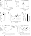
Figure 1.
Figure 1.
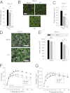
Acute lung injury (ALI) induces body weight loss, reduced food consumption, and muscle weakness. Bronchoalveolar lavage fluid (BALF) of ALI (intratracheal [IT]-LPS) and sham (IT-H2O) mice revealed an increase in total cell count (A) and total protein (B) that peaked between Days 3 and 4 in ALI mice and returned to baseline by Day 10. *P < 0.03 versus baseline values; n = 5–6. Based on these data and the accompanying histology of the lungs (see Figure E1), we refer to the period between Days 1 and 4 after IT-LPS as the “lung injury phase” and between Days 4 and 10 as the “lung resolution phase.” (C) ALI mice have an approximate 20% drop in body weight (BW) 3 days after IT-LPS. P < 0.001; n = 5. (D) Food consumption dramatically falls in ALI mice after IT-LPS (open triangles) compared with sham-operated mice (black circles). P < 0.01. (E) Pair-fed (PF) mice were used to control for the effects of reduced food consumption. These mice underwent a sham surgery (IT-H2O) and had food intake matched to that consumed by ALI mice on a daily basis. The data in the bar graphs show that cumulative food intake over the 10 days of lung injury and resolution was equal in PF and ALI mice. P < 0.01. (F) Whole body weight loss profiles were similar in PF (gray circles) and ALI mice (open triangles) but significantly different from changes in the body weights of sham mice (black circles) (analysis of variance, P < 0.001 for group differences), except for Day 3. §P < 0.03; n = 5. (G) Despite similar body weight losses, ALI mice had reduced grip strength compared with PF mice (analysis of variance, P < 0.001). Data are expressed as means ± SEM. *P < 0.001, ALI versus PF; P = 0.008, sham versus ALI; P = 0.014, sham versus PF. n = 8–10 in each group.
Figure 2.
Figure 2.
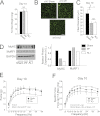
Acute lung injury (ALI) and pair-fed (PF) mice develop skeletal muscle atrophy. (A) Tibialis anterior (TA) muscle wet weights were decreased in PF and ALI mice by Day 3 post–intratracheal (IT)-LPS. *P < 0.003; n = 6. (B) Cross-sections of TA muscles from sham, ALI, and PF mice at Day 4 post–IT-LPS. Muscle fibers were identified by sarcolemmal staining for γ-laminin. Scale bar = 100 μm. (C) Mean fiber diameters of sham, PF, and ALI from Day 4 mice. *P < 0.003; n = 5. (D) Combined myosin ATPase histochemistry (alkaline preincubation) and γ-laminin staining enabled independent fiber size measurements of type I (light staining) and type II (dark staining) muscle fibers in the soleus. (E) Summary of mean muscle fiber diameters segregated by experimental group and muscle fiber type. Data are expressed as means ± SEM. §P < 0.03; P < 0.02. Scale bar as in (B). (F) Force-frequency curves for Day 4 sham, PF, and ALI hindlimb muscles showing reduced contractile force production in PF and ALI mice. (G) Specific force–frequency curves for Day 4 sham, PF, and ALI mice. Normalization of force to muscle mass for all three groups shows no difference in contractile performance.
Figure 3.
Figure 3.
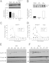
Myosin heavy chain (MHC) degradation, ubiquitination, and increased muscle atrogene expression in pair-fed (PF) and acute lung injury (ALI) mice. (A) Western blots showing sarcomeric MHC abundance in sham, PF, and ALI mice and MHC from ALI muscle ring finger-1 (MuRF1)−/− mice (top). Quantification of expression normalized to glyceraldehyde phosphate dehydrogenase (GAPDH) expression (bottom). (B) Western blot of extracts from sham, PF, and ALI muscles show a marked increase in high molecular weight (HMW) polyubiquinated proteins (top). Quantification of expression expressed in arbitrary units (bottom). Results of quantitative reverse transcriptase polymerase chain reaction measurements of MuRF1 (C) and atrogin1 (D) mRNA expression in muscles from PF and ALI mice versus expression in sham mice. *P < 0.02; n = 5–8. (E and F) Time course of MuRF1 and atrogin1 protein expression in PF and ALI mice. PF mice exhibit a relatively weak expression of MuRF1 proteins, whereas up-regulation of MuRF1 protein in ALI is much stronger. Neither PF nor ALI muscles exhibit any change in atrogin1 protein levels at any time and under any condition tested. *Possible processing intermediates of MuRF1.
Figure 4.
Figure 4.
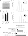
Muscle wasting in muscle ring finger-1 (MuRF1)−/− mice and increased nuclear factor (NF)-κB activity in acute lung injury (ALI) wild-type (WT) mouse muscles. (A) Western blot of MuRF1 protein expression shows up-regulation in ALI MuRF1 WT but no up-regulation in ALI MuRF1−/− mouse muscles. GAPDH = glyceraldehyde phosphate dehydrogenase; PF = pair fed. (B) Mean fiber diameters for WT and MuRF1−/− tibialis anterior (TA) muscles show a moderate increase in mean muscle fiber diameters in sham control MuRF1−/− mice. Exposure to ALI markedly reduces mean muscle fiber diameter in WT muscles mice but only a slight reduction in the MuRF1−/− muscle. *P = 0.004; P = 0.007; #P = 0.03. (C) Histograms of the fiber distribution of sham and ALI-treated MuRF1+/+ (WT) muscles reveal a large shift to the left (smaller size). (D) Histograms of the fiber distribution of sham and ALI-treated MuRF1−/− (knockout) muscles show only a small shift to the left and smaller fiber diameters. (E) Assessment of ALI-induced muscle transcriptional activity was measured by electroporating plasmids in which firefly luciferase transcription was controlled by transcriptional response elements specific for glucorticoid signaling (4XGRE-luc), FOXO signaling (3XFBE-luc), and NF-κB signaling (3XNf-κB-luc). Only increases in FBE- and NF-κB–luciferase activity is significantly up-regulated in ALI. (F) Luciferase reporter for MuRF1 transcriptional activity containing either no NF-κB binding elements (500 MuRF1-luc) or multiple NF-κB binding sites (5,000 MuRF-luc).
Figure 5.
Figure 5.
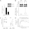
Muscle ring finger-1 (MuRF1) miRNA-based short hairpin (sh) RNAs effectively blocks MuRF1 protein expression in pair-fed (PF) and acute lung injury (ALI) mice but increases myosin heavy chain (MHC) content only in acute lung injury (ALI) mice. (A) Immunoblots of MuRF1 and glyceraldehyde phosphate dehydrogenase (GAPDH) from muscles transduced with Emerald GFP (EmGFP)-Neg control (NEG) shRNA or EmGFP-MuRF1 shRNA show that MuRF1 expression is reduced by approximately 70% in MuRF1 shRNA muscles. *P < 0.008. (B) Knockdown of MuRF1 protein by the EmGFP-MuRF1shRNA plasmid increased MHC content in ALI mice but had no effect on MHC levels in PF mice. Dotted line in B represents MHC level in control (EmGFP-Neg shRNA) muscles. P = 0.03. (C) Differences in tibialis anterior (TA) muscle mass in muscle transduced with EmGFP-MuRF1 shRNA, with results expressed with reference to EmGFP-Neg shRNA. (D) In situ force–frequency curves for muscles transduced with EmGFP-Neg and EmGFP-MuRF shRNAs and analyzed at Day 10. Data show an increase in force production by MuRF1 shRNA versus NEG shRNA. P = 0.023.
Figure 6.
Figure 6.
Muscle ring finger-1 (MuRF1) regulates muscle atrophy in acute lung injury (ALI) mice. Emerald GFP (EmGFP)-Neg shRNA or EmGFP-MuRF1 shRNAs were electroporated into the tibialis anterior (TA) muscles. One week later, mice underwent sham, pair-fed (PF), or ALI treatments. (A) Muscles were fixed at harvest and the sarcolemma stained with an antibody to dystrophin (red) to measure diameters of fibers expressing EmGFP. Scale = 100 μM. (B) Fiber histograms of sham, PF, and ALI mice treated with EMGFP-MuRF1 shRNA or EmGFP-Neg shRNAs. (C) The same technique was used to electroporate EmGFP-atrogin1 shRNA into the TA of ALI mice. (D) Mean fiber diameters of sham, PF, and ALI mice treated with MuRF1 or atrogin1 shRNAs. Fiber diameter is restored to sham conditions in ALI mice treated with MuRF1 shRNA but not atrogin1 shRNA. Data are expressed as means ± SEM. *P < 0.006; n = 5 in each group.
Figure 7.
Figure 7.
Persistence of muscle loss after complete resolution of lung injury. (A) Tibialis anterior (TA) wet weights at Day 10 in sham, pair-fed (PF), and acute lung injury (ALI) TA muscles are not significantly different. (B) Cryosections of γ-laminin–stained muscle sections from Day 10 sham, PF, and ALI mice. (C) Mean muscle fiber analysis of Day 10 muscles shows similar reductions in PF and ALI diameters compared with sham controls. P < 0.001; n = 5. (D) Representative Western blots of myosin heavy chain (MyHC) and muscle ring finger-1 (MuRF1) protein expression shows that MyHCs remain reduced at Day 10, whereas MuRF1 protein expression is reduced in all groups (Figures 3E and 3F show a time course of MuRF1 protein expression from Days 0–10). GAPDH = glyceraldehyde phosphate dehydrogenase. (E) Force–frequency curves for Day 10 muscles show that ALI sham and PF curves are not significantly different for each other but are both significantly different from ALI muscle (P = 0.01). (F) Specific force–frequency curves at Day 10 show a significant difference between PF and ALI muscles (P = 0.013).

Comment in

References

    1. Ware LB, Matthay MA. The acute respiratory distress syndrome. N Engl J Med 2000;342:1334–1349 - PubMed
    1. Rubenfeld GD, Caldwell E, Peabody E, Weaver J, Martin DP, Neff M, Stern EJ, Hudson LD. Incidence and outcomes of acute lung injury. N Engl J Med 2005;353:1685–1693 - PubMed
    1. Ali NA, O'Brien JM, Jr, Hoffmann SP, Phillips G, Garland A, Finley JC, Almoosa K, Hejal R, Wolf KM, Lemeshow S, et al. ; Midwest Critical Care Consortium Acquired weakness, handgrip strength, and mortality in critically ill patients. Am J Respir Crit Care Med 2008;178:261–268 - PubMed
    1. Fletcher SN, Kennedy DD, Ghosh IR, Misra VP, Kiff K, Coakley JH, Hinds CJ. Persistent neuromuscular and neurophysiologic abnormalities in long-term survivors of prolonged critical illness. Crit Care Med 2003;31:1012–1016 - PubMed
    1. Herridge MS, Cheung AM, Tansey CM, Matte-Martyn A, Diaz-Granados N, Al-Saidi F, Cooper AB, Guest CB, Mazer CD, Mehta S, et al. Canadian Critical Care Trials Group One-year outcomes in survivors of the acute respiratory distress syndrome. N Engl J Med 2003;348:683–693 - PubMed

Publication types

MeSH terms