Comparative Study
. 2012 Apr 15;185(8):851-61.
doi: 10.1164/rccm.201106-1152OC. Epub 2012 Feb 3.

Activation of mitochondrial biogenesis by heme oxygenase-1-mediated NF-E2-related factor-2 induction rescues mice from lethal Staphylococcus aureus sepsis

Affiliations
Comparative Study

Activation of mitochondrial biogenesis by heme oxygenase-1-mediated NF-E2-related factor-2 induction rescues mice from lethal Staphylococcus aureus sepsis

Nancy Chou MacGarvey et al. Am J Respir Crit Care Med. .

Abstract

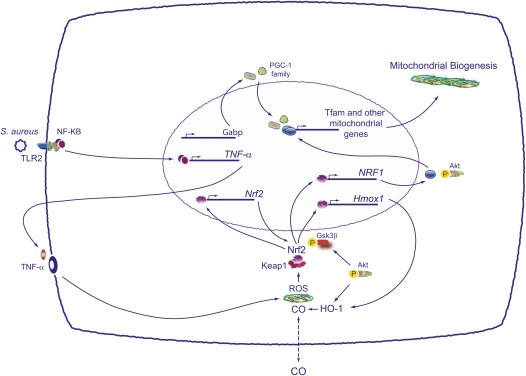
Rationale: Mitochondrial damage is an important component of multiple organ failure syndrome, a highly lethal complication of severe sepsis that lacks specific therapy. Mitochondrial quality control is regulated in part by the heme oxygenase-1 (HO-1; Hmox1) system through the redox-regulated NF-E2-related factor-2 (Nrf2) transcription factor, but its role in mitochondrial biogenesis in Staphylococcus aureus sepsis is unknown.

Objectives: To test the hypothesis that Nrf2-dependent up-regulation of the HO-1/carbon monoxide (CO) system would preserve mitochondrial biogenesis and rescue mice from lethal S. aureus sepsis.

Methods: A controlled murine S. aureus peritonitis model with and without inhaled CO was examined for HO-1 and Nrf2 regulation of mitochondrial biogenesis and the resolution of hepatic mitochondrial damage.

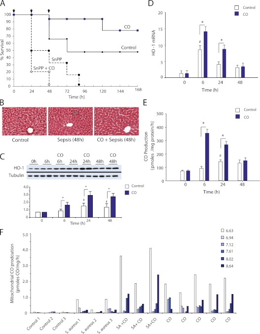
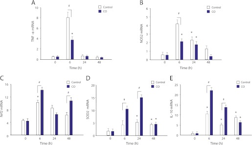
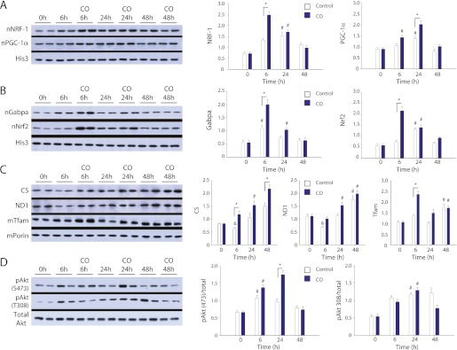
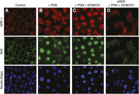
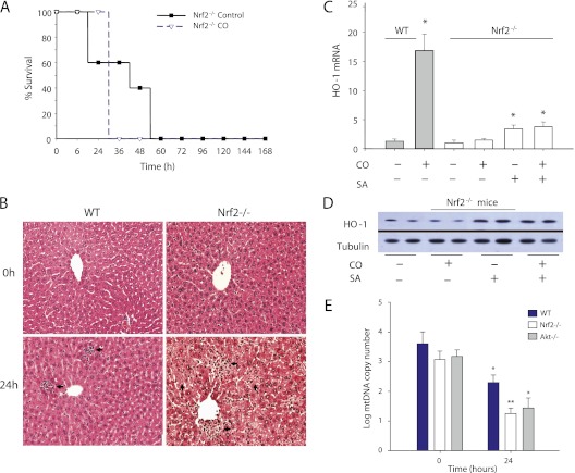
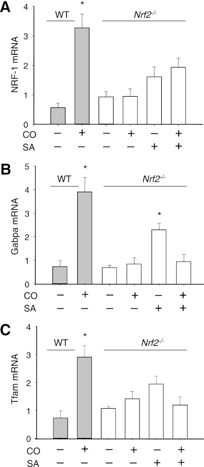
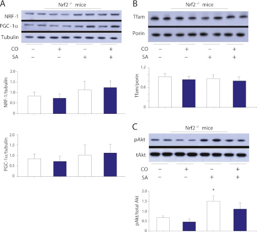
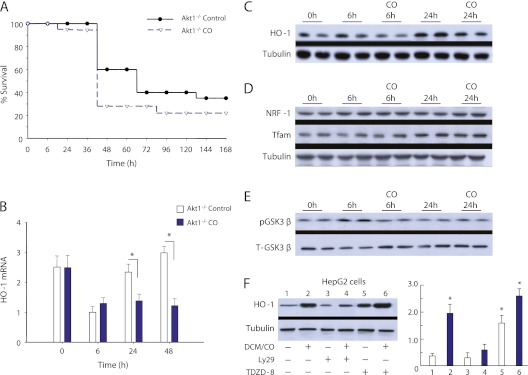
Measurements and main results: Sepsis survival was significantly enhanced using inhaled CO (250 ppm once-daily for 1 h), and linked mechanistically to Hmox1 induction and mitochondrial HO activity through Nrf2 transcriptional and Akt kinase activity. HO-1/CO stimulated Nrf2-dependent gene expression and nuclear accumulation of nuclear respiratory factor-1, -2α (Gabpa), and peroxisome proliferator-activated receptor gamma coactivator-1α; increased mitochondrial transcription factor-A and citrate synthase protein levels; and augmented mtDNA copy number. CO enhanced antiinflammatory IL-10 and reduced proinflammatory tumor necrosis factor-α production. By contrast, Nrf2(-/-) and Akt1(-/-) mice lacked CO induction of Hmox1 and mitochondrial biogenesis, and CO rescued neither strain from S. aureus sepsis.

Conclusions: We identify an inducible Nrf2/HO-1 regulatory cycle for mitochondrial biogenesis that is prosurvival and counter-inflammatory in sepsis, and describe targeted induction of mitochondrial biogenesis as a potential multiple organ failure therapy.


Figures

Figure 1.
Figure 1.
Staphylococcus aureus peritonitis and hepatic heme oxygenase-1 (HO-1; Hmox1) gene, HO-1 protein, and HO enzyme activity in wild-type (WT) mice. (A) Survival of WT sepsis-control or sepsis–carbon monoxide (CO) mice (arrows) after implantation of fibrin clots containing 5 × 10⁷ cfu of S. aureus. Data are pooled from experiments of 12 mice per group done in duplicate. Inhaled CO is protective in S. aureus sepsis in WT mice (P = 0.014 by Pearson chi square); however, CO protection is lost when HO is inhibited with SnPP. (B) Histologic sections of the mouse liver stained with hematoxylin and eosin demonstrate increased inflammatory foci after S. aureus peritonitis and protection after intervention with CO. (C) Western analysis for HO-1 protein in sepsis-control and sepsis-CO mice showing increases in the hepatic protein level in sepsis-control and sepsis-CO mice (densitometry bars are mean ± SD for n = 4 per group and time; #P < 0.05 vs. time 0; *P < 0.05, vs. sepsis-control). (D) Real-time reverse transcriptase polymerase chain reaction for hepatic HO-1 mRNA in WT sepsis-control and sepsis-CO mice. HO-1 mRNA is significantly increased by CO. Bars are means ± SD of duplicates in three experiments (#P < 0.05 vs. time 0; *P < 0.05 vs. sepsis-control group at the same time point). (E) Hepatic HO enzyme activity in WT sepsis-control and sepsis-CO mice. HO activity peaks at 24 hours in sepsis-control and at 6 hours in sepsis-CO mice. Bars are mean ± SD of duplicates in three experiments (#P < 0.05 vs. time 0; *P < 0.05, vs. sepsis-control). (F) Distribution of liver mitochondrial HO activity (per microgram mitochondrial protein) as a function of mitochondrial pHi in control mice and after 24 hours in sepsis-control, sepsis-CO, and CO-only mice. HO enzyme activity was enriched in sepsis and highest in mitochondria at the extremes of pHi, implying activation of heme degradation in these subpopulations.
Figure 2.
Figure 2.
Hepatic tumor necrosis factor (TNF)-α, nitric oxide synthase (NOS)-2, IL-10, and NF-E2–related factor-2 (Nrf2) antioxidant responses by real-time reverse transcriptase polymerase chain reaction during Staphylococcus aureus sepsis. (A) TNF-α mRNA up-regulation in sepsis-control mice is attenuated in sepsis–carbon monoxide (CO) mice (*P < 0.05 vs. time 0; #P < 0.05 vs. time 0 and sepsis-CO). (B) NOS2 mRNA is induced in sepsis-control mice by 6 hours, but is limited by inhaled CO (A and B: *P < 0.05 vs. time 0; #P < 0.05 vs. time 0 and with CO). (C) Nrf2 mRNA by real-time reverse transcriptase polymerase chain reaction is increased in sepsis-control mice by 6 hours, and expression of the gene is enhanced by CO inhalation. (D) Increased superoxide dismutase 2 mRNA levels in sepsis-control mice are further increased in sepsis-CO mice. (E) Increased IL-10 mRNA levels in sepsis-control mice are further increased in sepsis-CO mice (C, D, and E: *P < 0.05 vs. time 0; #P < 0.05 vs. time 0 and sepsis-control mice).
Figure 3.
Figure 3.
Transcriptional up-regulation of mitochondrial biogenesis and protection of mtDNA copy number in Staphylococcus aureus sepsis. (A) Hepatic nuclear respiratory factor (NRF)-1 expression by real-time reverse transcriptase polymerase chain reaction increases in sepsis-control mice, reaching a maximum at 24 hours, with further increases in sepsis–carbon monoxide (CO) mice. (B) Hepatic up-regulation of Gabpa (NRF-2α) mRNA in sepsis-control mice parallels NRF-1 and further increases in sepsis-CO mice. (C) The peroxisome proliferator-activated receptor gamma coactivator (PGC)-1α coactivator is up-regulated by 6 hours of sepsis and further increases in sepsis-CO mice. (D) The PGC-1 family member PGC-1–related coactivator increases in sepsis-control mice and further increases in sepsis-CO mice. (E) Hepatic transcription factor-A (Tfam) mRNA level increases in sepsis-control mice and increases further in sepsis-CO mice. (A–E: #P < 0.05 vs. time 0; *P < 0.05 vs. time 0 and sepsis-control for n = 4 per time per group). (F) Log hepatic mitochondrial DNA copy number in sepsis-control and sepsis-CO mice. Inhaled CO reverses the decline in mtDNA copy number in sepsis (n = 4 per time per group; #P < 0.05 less than time 0 control; *P < 0.05 higher than the respective sepsis-control group).
Figure 4.
Figure 4.
Up-regulation of mitochondrial biogenesis proteins during Staphylococcus aureus sepsis in wild-type mice by Western blot analysis. (A) Hepatic nuclear respiratory factor (NRF)-1 and peroxisome proliferator-activated receptor gamma coactivator-1α nuclear proteins increase in sepsis-control mice and increase further with carbon-monoxide (CO) (His3 is a nuclear reference protein). (B) Western analysis of hepatic Gabpa (NRF-2α) showing enhanced nuclear protein levels in sepsis-control and sepsis-CO mice. Nuclear NF-E2–related factor-2 (Nrf2) protein in sepsis-control and sepsis-CO mice is shown from the same nuclear samples compared with His3. For panels A and B, the densitometry confirms significant up-regulation of all four proteins by 24 hours in sepsis-control mice and further increases in sepsis-CO mice (for the densitometry, n = 4; #P < 0.05 vs. time 0; *P < 0.05 for the CO effect). (C) To verify the induction of mitochondrial biogenesis, we checked three mitochondrial marker proteins (nuclear-encoded citrate synthase [CS], mtDNA-encoded Complex I subunit NADH dehydrogenase 1, and the imported mitochondrial transcription factor transcription factor-A [Tfam]) (for the densitometry n = 4; # P < 0.05 above time 0 control; &P < 0.05 below time 0; *P < 0.05 for the CO effect). (D) Akt phosphorylation in sepsis-control mice is shown to increase at S473 and T308, but CO only enhances S473 phosphorylation (for densitometry, n = 4; #P < 0.05 vs. time 0; *P < 0.05 for the CO effect).
Figure 5.
Figure 5.
Nuclear NF-E2–related factor-2 (Nrf2) and nuclear respiratory factor (NRF)-1 accumulation after Staphylococcus aureus peptidoglycan (PGN) or dichloromethane (DCM)/carbon monoxide (CO) administration in HepG2 cells. (A) HepG2 cells were examined by confocal microscopy for the localization and expression of NRF-1 (red) and Nrf2 (green). Before PGN, both transcription factors are detectable only in the cytoplasm. Nuclear staining with Dapi (blue) is shown in the bottom panels of the columns. (B) After PGN exposure, both transcription factors accumulate in cell nuclei. (C) After exposure to PGN plus DCM/CO, intense homogeneous nuclear staining is noted for Nrf2 and NRF-1. (D) Nrf2 siRNA (siNrf2) blocks the effects of PGN plus DCM/CO on the nuclear accumulation of Nrf2 and NRF-1. Scrambled siRNA had no effect (no images shown).
Figure 6.
Figure 6.
NF-E2–related factor-2 (Nrf2) deletion and Staphylococcus aureus sepsis. (A) Nrf2/ mice were implanted with fibrin clots containing S. aureus (5 × 10⁶ cfu). The mice in the sepsis-control group all died by 54 hours and carbon monoxide (CO) offered no advantage (100% mortality by ∼ 36 h; P = NS; n = 12 mice per group). (B) Hematoxylin and eosin–stained liver sections in wild-type (WT) (left) and Nrf2/ mice (right) at 0 and 24 hours after S. aureus inoculation (5 × 10⁶ cfu). Extensive cell vacuolization, autolysis, and cell death are present in Nrf2/ mouse liver. Horizontal arrows indicate inflammatory foci after sepsis, which are larger and more numerous in Nrf2/ mice. Vertical arrows indicate foci of necrosis or apoptosis. (C) Heme oxygenase (HO)-1 mRNA levels in WT and Nrf2/ mice. The WT response to CO is a positive control. In Nrf2/ mice, S. aureus sepsis but not CO induces HO-1 mRNA expression (*P < 0.05 vs. sepsis-CO). (D) Hepatic HO-1 protein in Nrf2/ mice by Western blot. S. aureus increases HO-1 protein, whereas CO does not. (E) Loss of hepatic mtDNA copy number in Nrf2/ and Akt1−/− mice compared with WT mice at 24 hours of sepsis (*P < 0.05 compared with WT at time 0; **P < 0.05 compared with WT at 0 and 24 h). Note the higher S. aureus inoculation dose in WT mice. SA = S. aureus.
Figure 7.
Figure 7.
Transcriptional regulators of mitochondrial biogenesis in NF-E2–related factor-2 (Nrf2)/ mice during Staphylococcus aureus sepsis. (A) Liver nuclear respiratory factor (NRF)-1 mRNA levels by real-time reverse transcriptase polymerase chain reaction increase less than twofold in Nrf2/ sepsis-control mice and there is no significant response to CO. CO response in wild-type (WT) mice is provided as a positive control. (B) Gabpa mRNA levels in Nrf2/ sepsis-control mice increase 2.5-fold, but there is no effect of CO. (C) Transcription factor-A (Tfam) mRNA expression does not increase in Nrf2/ sepsis-control or sepsis-CO mice. Values are means ± SD of duplicates of three experiments (*P < 0.05 vs. baseline control). SA = S. aureus.
Figure 8.
Figure 8.
Regulation of mitochondrial biogenesis regulatory proteins in NF-E2–related factor-2 (Nrf2)/ mice during Staphylococcus aureus sepsis. (A) Western blots of total hepatic nuclear respiratory factor (NRF)-1 and PGC-1α protein showing minimal increases in Nrf2/ sepsis-control mice, but none in carbon monoxide (CO) alone or in sepsis-CO mice (for densitometry, n = 4 per group). (B) Transcription factor-A (Tfam) is unaffected in Nrf2/ sepsis-control, CO-only, or sepsis-CO mice (densitometry n = 4 per group). (C) In Nrf2/ mice, Akt (S473) is phosphorylated in Nrf2/ sepsis-control mice, but there are no effects of CO (densitometry n = 4 per group). SA = S. aureus.
Figure 9.
Figure 9.
Responses of Akt1/ mice to Staphylococcus aureus sepsis. (A) Survival in Akt1/ mice implanted with 5 × 10⁶ cfu S. aureus was impaired compared with wild-type mice (Figure 1A) and did not improve with carbon monoxide (CO) administration. Seven-day mortality was 78% in sepsis-CO compared with 64% in sepsis-control mice (n = 12 per group; P = NS). (B) Real-time reverse transcriptase polymerase chain reaction for heme oxygenase (HO)-1 mRNA in Akt1/ mice, unlike wild-type, shows no increase in sepsis. CO did not increase HO-1 mRNA in Akt1/ mice, and mRNA levels are lower than sepsis-control mice at 24 and 48 hours (*P < 0.05 vs. CO). (C) Western blot showing increases in hepatic HO-1 protein levels in Akt1/ sepsis-control mice at 24 hours, but CO had no effect on HO-1. (D) Hepatic protein levels for nuclear respiratory factor (NRF)-1 and transcription factor-A (Tfam) did not increase in Akt/ sepsis-control or sepsis-CO mice. (E) Hepatic GSK3β phosphorylation increased in Akt/ sepsis-control mice only at 6 hours, and CO had no effect. (F) HepG2 cells incubated with dichloromethane (DCM)/CO demonstrate the effect of CO on HO-1 protein expression. HO-1 induction by DCM/CO was blocked by inhibition of Akt and enhanced by inhibition of Gsk3β (densitometry n = 3; *P < 0.05 compared with lane 1).
Figure 10.
Figure 10.
The heme oxygenase (HO)-1/carbon monoxide (CO) system and activation of mitochondrial biogenesis in Staphylococcus aureus sepsis. The diagram illustrates the redox cycle that activates NF-E2–related factor-2 (Nrf2) and the HO-1/CO system in response to S. aureus and to CO. Both the oxidative stress of innate immune hyperactivation and the CO intervention induce the mitochondrial biogenesis transcriptional program through Nrf2. The two known sites of Akt involvement are also indicated. Keap1 = Kelch-like ECH-associated protein 1; NF-κB = nuclear factor-κB; NRF1 = nuclear respiratory factor-1; PGC = peroxisome proliferator-activated receptor gamma coactivator; Tfam = transcription factor-A; ROS = reactive oxygen species; TLR = Toll-like receptor; TNF = tumor necrosis factor.

Comment in

References

    1. Dombrovskiy VY, Martin AA, Sunderram J, Paz HL. Rapid increase in hospitalization and mortality rates for severe sepsis in the United States: a trend analysis from 1993 to 2003. Crit Care Med 2007;35:1244–1250 - PubMed
    1. Martin GS, Mannino DM, Eaton S, Moss M. The epidemiology of sepsis in the United States from 1979 through 2000. N Engl J Med 2003;348:1546–1554 - PubMed
    1. Brealey D, Brand M, Hargreaves I, Heales S, Land J, Smolenski R, Davies NA, Cooper CE, Singer M. Association between mitochondrial dysfunction and severity and outcome of septic shock. Lancet 2002;360:219–223 - PubMed
    1. Vincent JL, Bruzzi de Carvalho F. Severity of illness. Semin Respir Crit Care Med 2010;31:31–38 - PubMed
    1. Bradley JR. TNF-mediated inflammatory disease. J Pathol 2008;214:149–160 - PubMed

Publication types

MeSH terms