. 2012 Jun;46(6):833-41.
doi: 10.1165/rcmb.2011-0230OC. Epub 2012 Feb 3.

Virus-infected alveolar epithelial cells direct neutrophil chemotaxis and inhibit their apoptosis

Affiliations

Virus-infected alveolar epithelial cells direct neutrophil chemotaxis and inhibit their apoptosis

Joanna P Rzepka et al. Am J Respir Cell Mol Biol. 2012 Jun.

Abstract

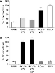
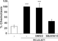
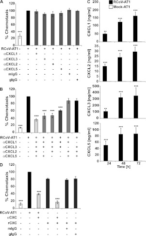
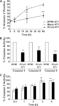
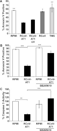
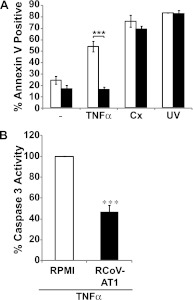
The alveolar epithelium is a critical target for pulmonary viruses and can produce proinflammatory cytokines and chemokines upon viral infection. However, the molecular interactions between virus-infected alveolar epithelial cells and inflammatory cells, including polymorphonuclear leukocytes (PMNs), have not been thoroughly characterized. Rat coronavirus (RCoV) is used as a model to study the immune response to viral infection in the lung of the natural host. We have developed an in vitro model to characterize the response of PMNs to RCoV-infected type I-like alveolar epithelial (AT1) cells, the primary target for RCoV infection in the alveoli. Multiple CXC chemokines that signal through CXCR2 were required for PMN chemotaxis toward medium from RCoV-infected AT1-like cells (RCoV-AT1). Furthermore, RCoV-AT1 inhibited spontaneous PMN apoptosis, including activation of effector caspase 3 and initiator caspases 8 and 9. Use of a selective inhibitor of CXCR2, SB265610, demonstrated that CXCR2 signaling was required for RCoV-AT1-mediated inhibition of PMN apoptosis. These data suggest that CXC chemokines produced by RCoV-infected AT1-like cells inhibit PMN apoptosis during infection. These studies provide new insight into the molecular mechanisms whereby alveolar epithelial cells direct the functions of PMNs during viral infection of the lung.


Figures

Figure 1.
Figure 1.
Chemotaxis of rat polymorphonuclear leukocytes (PMNs) toward medium from rat coronavirus (RCoV)-infected AT1-like cells. Rat PMNs were isolated from bone marrow and stained with calcein-AM, and the chemotaxis of 3 × 10⁴ PMNs was quantified by fluorescence intensity in a micro-chemotaxis plate. (A) RPMI, medium from uninfected (RPMI-AT1 and Mock-AT1) or RCoV-infected (RCoV-AT1) AT1-like cells, or FMLP were placed in the lower chamber of the chemotaxis plate. Fluorescence intensity data were normalized to FMLP. (B) RCoV-AT1, UV-irradiated RCoV-AT1, purified virus, and virus storage buffer (TMS) were placed in the lower chamber of the chemotaxis plate. Fluorescence intensity data were normalized to RCoV-AT1. Data are means ± SEM from at least three independent experiments with triplicate samples per experiment. Statistical significance versus RPMI: ***P < 0.001.
Figure 2.
Figure 2.
The role of CXCR2 signaling in PMN chemotaxis toward RCoV-AT1. Rat PMNs were isolated from bone marrow and stained with calcein-AM, and the chemotaxis of 3 × 10⁴ PMNs was quantified by fluorescence intensity in a micro-chemotaxis plate toward RPMI or RCoV-AT1 in the presence or absence of CXCR2 inhibitor (SB265610) or vehicle (DMSO). Fluorescence intensity data were normalized to RCoV-AT1. Data shown are the mean values ± SEM from three independent experiments with triplicate samples per experiment. Statistical significance versus RCoV-AT1 with SB265610: ***P < 0.001.
Figure 3.
Figure 3.
Chemotaxis of PMNs toward RCoV-AT1 in the presence of neutralizing antibodies to CXC chemokines. Rat PMNs were isolated from bone marrow and stained with calcein-AM, and the chemotaxis of 3 × 10⁴ PMNs toward RCoV-AT1 was quantified by fluorescence intensity in a micro-chemotaxis plate. RCoV-AT1 was incubated with the indicated antibodies before chemotaxis assay. Fluorescence intensity data were normalized to RCoV-AT1. (A) The ability of individual αCXC antibodies to neutralize chemotaxis toward RCoV-AT1 was evaluated. Mouse IgG (mIgG) was used as an isotype control for αCXCL2, -3, and -5 and goat IgG (gIgG) was used as an isotype control for αCXCL1. (B) Multiple antibodies were evaluated in combination. *** Statistical significance versus RCoV-AT1 (P < 0.001). (C) The concentrations of CXC chemokines in RCoV-AT1 compared with mock-AT1 at 24, 48, and 72 hours after inoculation were quantified by ELISA. Statistical significance versus mock-AT1: ** P < 0.01, *** P < 0.001. (D) Chemotaxis of PMNs toward recombinant CXC chemokines (rCXC) at equivalent concentrations as in RCoV-AT1 was quantified with or without the addition of neutralizing antibodies to the four chemokines (αCXC). *** Statistical significance versus RCoV-AT1 (P < 0.001). Data shown are means ± SEM from at least three independent experiments with triplicate samples per experiment.
Figure 4.
Figure 4.
RCoV-AT1–mediated inhibition of spontaneous PMN apoptosis. (A) PMNs were isolated from rat blood and incubated with supernatant medium from noninfected (RPMI-AT1), mock-infected (Mock-AT1), or RCoV-infected (RCoV-AT1) AT1-like cells and annexin V-FITC binding was analyzed by flow cytometry. (B) The activities of effector caspase 3 (after 6-h incubation), initiator caspases 8 and 9 (after 4-h incubation), and (C) caspase 2 (at indicated times) were quantified in PMNs incubated in RPMI or RCoV-AT1 using luminescent substrate assays. Luminescence intensities were normalized to PMNs incubated in RPMI. Data shown are means ± SEM from at least three independent experiments. Statistical significance versus (A) RPMI-AT1, (B) RPMI, or (C) RPMI at 0.5 h: *P < 0.05, **P < 0.01, and ***P < 0.001.
Figure 5.
Figure 5.
The role of viral particles and CXCR2 signaling in RCoV-AT1–mediated inhibition of spontaneous PMN apoptosis. PMNs were isolated from rat blood. (A) Apoptosis of PMNs incubated with UV-irradiated RCoV-AT1 or purified RCoV or virus storage buffer (TMS) for 24 hours was analyzed by annexin V-FITC binding and flow cytometry. Apoptosis of PMNs incubated with RCoV-AT1 with or without CXCR2 inhibitor (SB265610) was analyzed by (B) annexin V-FITC binding and (C) caspase 3 activity using a luminescent substrate assay. Luminescence data were normalized to PMNs incubated in RPMI. Data shown are means ± SEM from at least three independent experiments. Statistical significance versus RCoV-AT1: *P < 0.05, **P < 0.01, and ***P < 0.001.
Figure 6.
Figure 6.
RCoV-AT1–mediated inhibition of induced PMN apoptosis. PMNs were isolated from rat blood and preexposed to UV irradiation or incubated with TNF-α or cycloheximide (Cx) in RPMI or RCoV-AT1. (A) Annexin V-FITC binding was quantified by flow cytometry, and (B) caspase 3 activity was analyzed by luminescent substrate assay. Luminescence data were normalized to PMNs incubated in RPMI with TNF-α. Data shown are the mean values ± SEM from at least three independent experiments. Statistical significance as indicated (***P < 0.001).

References

    1. Shieh W-J, Blau DM, Denison AM, DeLeon-Carnes M, Adem P, Bhatnagar J, Sumner J, Liu L, Patel M, Batten B, et al. 2009 Pandemic Influenza A (H1N1): pathology and pathogenesis of 100 fatal cases in the United States. Am J Pathol 2010;177:166–175 - PMC - PubMed
    1. Johnson JE, Gonzales RA, Olson SJ, Wright PF, Graham BS. The histopathology of fatal untreated human respiratory syncytial virus infection. Mod Pathol 2007;20:108–119 - PubMed
    1. Nicholls JM, Butany J, Poon LL, Chan KH, Beh SL, Poutanen S, Peiris JS, Wong M. Time course and cellular localization of SARS-CoV nucleoprotein and RNA in lungs from fatal cases of SARS. PLoS Med 2006;3:e27. - PMC - PubMed
    1. Uiprasertkul M, Kitphati R, Puthavathana P, Kriwong R, Kongchanagul A, Ungchusak K, Angkasekwinai S, Chokephaibulkit K, Srisook K, Vanprapar N, et al. Apoptosis and pathogenesis of avian influenza a (H5N1) virus in humans. Emerg Infect Dis 2007;13:708–712 - PMC - PubMed
    1. Roberts A, Deming D, Paddock CD, Cheng A, Yount B, Vogel L, Herman BD, Sheahan T, Heise M, Genrich GL, et al. A mouse-adapted SARS-coronavirus causes disease and mortality in balb/c mice. PLoS Pathog 2007;3:e5. - PMC - PubMed

Publication types