

Anatomic considerations for central venous cannulation
- PMID: 22312225
- PMCID: PMC3270925
- DOI: 10.2147/RMHP.S10383
Anatomic considerations for central venous cannulation
Abstract
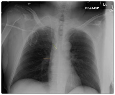
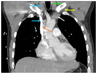
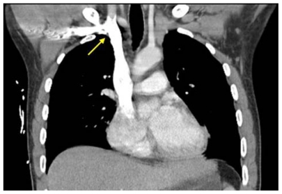
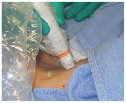
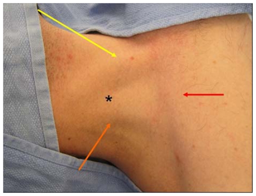
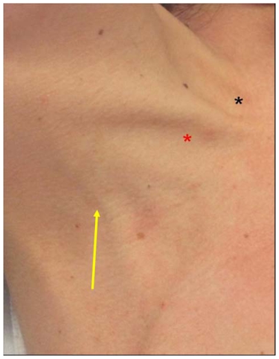
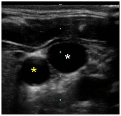
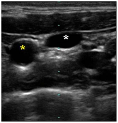
Central venous cannulation is a commonly performed procedure which facilitates resuscitation, nutritional support, and long-term vascular access. Mechanical complications most often occur during insertion and are intimately related to the anatomic relationship of the central veins. Working knowledge of surface and deep anatomy minimizes complications. Use of surface anatomic landmarks to orient the deep course of cannulating needle tracts appropriately comprises the crux of complication avoidance. The authors describe use of surface landmarks to facilitate safe placement of internal jugular, subclavian, and femoral venous catheters. The role of real-time sonography as a safety-enhancing adjunct is reviewed.
Keywords: Internal jugular vein; cannulation; ultrasound; venipuncture.
Figures
References
-
- McGee DC, Gould MK. Preventing complications of central venous catheterization. N Engl J Med. 2003;348:1123–1133. - PubMed
-
- Ellis H. The clinical anatomy of the great veins of the neck. Br J Hosp Med. 2010;71(2):M20–M21. - PubMed
-
- Boon JM, Van Schoor AN, Abrahams PH, Meiring JH, Welch T, Shanahan D. Central venous catheterization – An anatomical review of a clinical skill – Part 1: Subclavian vein via the infraclavicular approach. Clin Anat. 2007;20:602–611. - PubMed
-
- Boon JM, Van Schoor AN, Abrahams PH, Meiring JH, Welch T. Central venous catheterization – An anatomical review of a clinic skill, Part 2: Internal jugular vein via the supraclavicular approach. Clin Anat. 2008;21:15–22. - PubMed
-
- Maki DG, Ringer M, Alvarado CJ. Prospective randomized trial of povidone-iodine, alcohol, and chlorhexidine for prevention of infection associated with central venous and arterial catheters. Lancet. 1991;338:339–343. - PubMed
LinkOut - more resources
Full Text Sources
Other Literature Sources
