. 2012;7(2):e31302.
doi: 10.1371/journal.pone.0031302. Epub 2012 Feb 1.

Trans-synaptic spread of tau pathology in vivo

Affiliations

Trans-synaptic spread of tau pathology in vivo

Li Liu et al. PLoS One. 2012.

Abstract

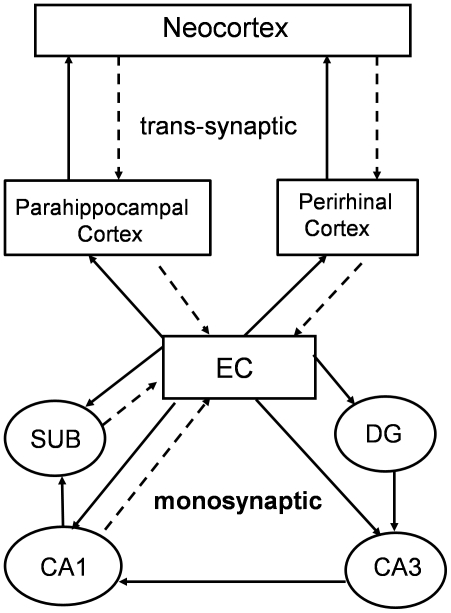
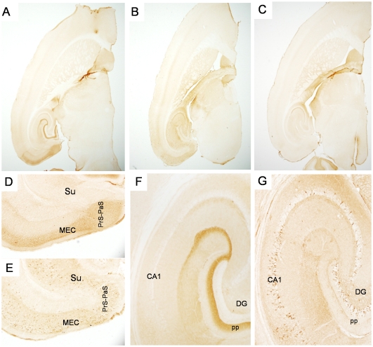
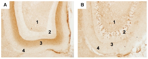
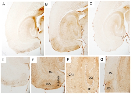
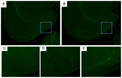
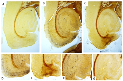
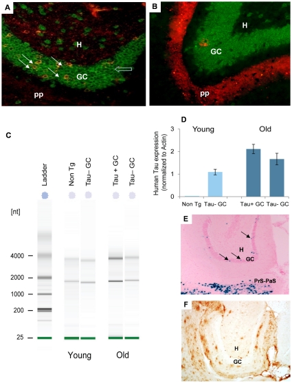
Tauopathy in the brain of patients with Alzheimer's disease starts in the entorhinal cortex (EC) and spreads anatomically in a defined pattern. To test whether pathology initiating in the EC spreads through the brain along synaptically connected circuits, we have generated a transgenic mouse model that differentially expresses pathological human tau in the EC and we have examined the distribution of tau pathology at different timepoints. In relatively young mice (10-11 months old), human tau was present in some cell bodies, but it was mostly observed in axons within the superficial layers of the medial and lateral EC, and at the terminal zones of the perforant pathway. In old mice (>22 months old), intense human tau immunoreactivity was readily detected not only in neurons in the superficial layers of the EC, but also in the subiculum, a substantial number of hippocampal pyramidal neurons especially in CA1, and in dentate gyrus granule cells. Scattered immunoreactive neurons were also seen in the deeper layers of the EC and in perirhinal and secondary somatosensory cortex. Immunoreactivity with the conformation-specific tau antibody MC1 correlated with the accumulation of argyrophilic material seen in old, but not young mice. In old mice, axonal human tau immunoreactivity, especially at the endzones of the perforant pathway, was greatly reduced. Relocalization of tau from axons to somatodendritic compartments and propagation of tauopathy to regions outside of the EC correlated with mature tangle formation in neurons in the EC as revealed by thioflavin-S staining. Our data demonstrate propagation of pathology from the EC and support a trans-synaptic mechanism of spread along anatomically connected networks, between connected and vulnerable neurons. In general, the mouse recapitulates the tauopathy that defines the early stages of AD and provides a model for testing mechanisms and functional outcomes associated with disease progression.


Conflict of interest statement

Competing Interests: The authors have declared that no competing interests exist.

Figures

Figure 1
Figure 1. Monosynaptic and trans-synaptic cortico-hippocampal and cortico-cortico connections.
Solid lines indicate projections radiating out from the EC, dotted lines indicate projections to the EC. Monosynaptically connected regions are connected across one synapse. Trans-synaptic regions are separated by more than one synapse.
Figure 2
Figure 2. Progressive spread of tauopathy in NT mice identified by antibody MC1.
Fig. 2A shows tau immunolabeled with the human tau specific, conformational antibody MC1 in a young NT mouse at low power, and higher power (Figs. 2D, G). Fig. 2B shows MC1 immunolabeling in an old NT mouse at low power, and higher power (Figs. 2E, H). Fig. 2F shows high power image of cells immunolabeled with MC1 within the MEC. Old NT mice show extensive accumulation of human tau in cell bodies in the EC and subiculum (Fig. 2H), and in synaptically connected areas in the hippocampus and neocortex (Fig. 2E). Fig. 2I shows accumulation of human tau in neurons of the perirhinal cortex and into the parietal region in the old NT mouse. Note the lack of neurite staining in the perirhinal cortex compared to the LEC. Fig. 2C shows lack of immunolabeling with the human specific antibody in an old, littermate control mouse (single transgenic tau responder mouse, no tTA) except for the non-specific staining of the fornix that was seen with all antibodies. MEC = medial entorinal cortex, LEC = lateral entorhinal cortex, Pe = perirhinal cortex, Par = parietal cortex, DG = dentate gyrus, CA1, CA3 = CA fields of hippocampus, Su = subiculum, Prp-PaS = pre-parasubiculum, pp = perforant pathway endzone. Figs. 2A–C magnification = 2×, Figs. 2D, E magnification = 4×, Figs. 2G–I magnification = 10×. Fig. 2F magnification = 40×.
Figure 3
Figure 3. Progressive spread of tauopathy in NT mice identified by antibody CP27.
Fig. 3 shows mice immunolabeled with CP27 (total human tau) at low power in a young NT mouse (Fig. 3A), an old NT mouse (Fig. 3B) or a control mouse (Fig. 3C). Higher power images from the young NT mouse (Figs. 3D, F), old NT mouse (Figs. 3E, G) with antibody CP27. CP27 is one of several antibodies that show non-specific staining of mossy fibers in the control mouse. Figs. 3D and E show the EC and subiculum whereas Figs. 3F and G show the CA and DG regions of the hippocampus. Figs. 3A–C magnification = 2×, Figs. 3D–G magnification = 10×.
Figure 4
Figure 4. Progressive spread of tauopathy to monosynaptically connected regions of the hippocampus.
Young NT mice (Fig. 4A) show accumulation of human tau immunolabeled with CP27 predominately in the endzones of the perforant pathway that terminate in the middle third of the molecular layer of the DG (area 3). Terminals from neurons in the LEC terminating in the outer third of the molecular layer are shown in area 4. Human tau was also seen in cells in the hilus (area 1). Granule cell layers of the DG (area 2) did not accumulate human tau at this age. Old NT mice (Fig. 4B) show accumulation of human tau in cell bodies in the granule cells of the DG (area 2). Increased accumulation of human tau is seen in layers 1, 2 and 4 but the perforant pathway endzone in layer 3 was significantly depleted of tau. Magnification = 20×.
Figure 5
Figure 5. Progressive spread of tauopathy in NT mice identified by antibody AT8.
Fig. 5 shows mouse brain tissue immunolabeled with AT8 (phospho-tau S202/205) at low power in a young NT mouse (Fig. 5A), an old NT mouse (Fig. 5B) or a control mouse (Fig. 5C). Higher power images from the young NT mouse (Fig. 5D), or old NT mouse (Figs. 5E–G) with antibody AT8. Figs. 5D and E show the EC and subiculum whereas 5F shows the CA and DG regions of the hippocampus. Fig. 5G shows the boundary between the LEC and the perirhinal cortex in the old NT mouse. Note the scarcity of neurite staining in the perirhinal cortex compared to the LEC. Figs. 5A–C magnification = 4×, Figs. 5D–G magnification = 10×.
Figure 6
Figure 6. Mature, filamentous neurofibrillary tangle formation in the EC of old NT mice.
Fig. 6A shows lack of thioS staining of neurons in the EC of an old control mouse. The boxed area is shown in the high power image in Fig. 6C. Fig. 6B shows thioS positive neurons in the EC of an old NT mouse. The boxed area is shown in the high power image in Fig. 6E. Fig. 6D shows lack of thioS staining in the same region of the young NT mouse. Figs. 6A and B magnification = 10×, Figs. 6C–E magnification = 20×.
Figure 7
Figure 7. Silver staining in NT mice.
Fig. 7A shows a lower power image of argyrophilic material in the hippocampus and cortex of a young NT mouse (Fig. 7A), an old NT mouse (Fig. 7B) and a control mouse (age matched to the old NT mouse) (Fig. 7C). Higher power images from the EC and subiculum of the old NT mouse (Fig. 7D) or control mouse (Fig. 7E). Figs. 7F and G show high power images of argyrophilic neurons in the hippocampus of the old NT mouse (Fig. 7F) or control mouse (Fig. 7G). Note the relatively abundant, dense staining in EC, subiculum and CA regions compared to the faint staining in the DG, which mirrors the staining pattern with MC1. Figs. A, B and C magnification = 4×, Figs. 7D–G magnification = 10×.
Figure 8
Figure 8. LCM-isolation of cell populations and assessment of mRNA levels by qRT-PCR.
Fig. 8A shows high power images of frozen sections from an old NT mouse double-immunolabeled with antibodies recognizing the neuronal marker NeuN (green) and human tau CP27 (red), before LCM capture of dentate gyrus granule cells (GCs). Co-localization between CP27 and the neuronal marker NeuN indicates that expression of human tau was neuronal in origin and glial expression was negligible, or absent. Human tau positive neurons (Tau+ GC, yellow cells, solid white arrow) and tau negative neurons (Tau− GC, green cells, open blue arrow) were isolated. Fig. 8B shows the GC layer from a young mouse. Granule cell neurons in the DG were not immunoreactive for human tau at this age therefore only green neurons (Tau− GCs) in this region were isolated by LCM. Fig. 8C shows a gel image from the Agilent Bioanalyzer of RNA extracted from LCM isolated cells. Fig. 8D shows human tau expression in LCM isolated non Tg and Tau− GC of a young animal and in Tau+ GC and Tau− GC neurons from an old NT mice. The relative expression changes (2−ΔΔCT) between the cell types were calculated after normalization to β-actin expression. Error bars represent the SD for triplicates of each RT-cDNA sample used for qPCR. One old and one young mouse are represented in Fig. 8D; analyses were performed on cells from three individual old mice, and two young mice. Fig. 8E shows the DG of the neuropsin-tTA-lacZ reporter gene mouse (3 months of age) with GCs expressing the responder indicated (arrows). Fig. 8F shows the DG of a neuropsin-tTA-APP mouse with the DG GCs indicated. Mice were 22 months of age; two mice were examined. Despite the presence of extensive plaque pathology in adjacent layers, no APP/Aβ staining was seen in the DG GCs from this mouse line. Figs. 8A, B magnification = 20×. pp = perforant pathway endzone, H = hilus, GC = dentate gyrus granule cells.

References

    1. Braak H, Braak E. Neuropathological stageing of Alzheimer-related changes. Acta Neuropathol. 1991;82:239–259. - PubMed
    1. van Groen T, Miettinen P, Kadish I. The entorhinal cortex of the mouse: organization of the projection to the hippocampal formation. Hippocampus. 2003;13:133–149. - PubMed
    1. Witter MP, Wouterlood FG, Naber PA, Van Haeften T. Anatomical organization of the parahippocampal-hippocampal network. Ann N Y Acad Sci. 2000;911:1–24. - PubMed
    1. Braak H, Del Tredici K. Alzheimer's pathogenesis: is there neuron-to-neuron propagation? Acta Neuropathol. 2011;121:589–595. - PubMed
    1. Blaizot X, Meguro K, Millien I, Baron JC, Chavoix C. Correlations between visual recognition memory and neocortical and hippocampal glucose metabolism after bilateral rhinal cortex lesions in the baboon: implications for Alzheimer's disease. J Neurosci. 2002;22:9166–9170. - PMC - PubMed

Publication types