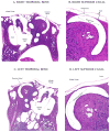
Histopathology of the temporal bone in a case of superior canal dehiscence syndrome
- PMID: 22312921
- PMCID: PMC3690372
- DOI: 10.1177/000348941212100102
Histopathology of the temporal bone in a case of superior canal dehiscence syndrome
Abstract
Objectives: We describe the histopathologic findings in the temporal bones of a patient who had, during life, received a diagnosis of superior canal dehiscence (SCD) syndrome.
Methods: The patient was found to have SCD syndrome at 59 years of age. She became a temporal bone donor, and died of unrelated causes at 62 years of age. Both temporal bones were prepared in celloidin and examined by light microscopy.
Results: The patient developed bilateral aural fullness, pulsatile tinnitus, and difficulty tolerating loud noises after minor head trauma at 53 years of age. The symptoms were worse on the right. She also had Valsalva-induced dizziness and eye movements, as well as sound-induced dizziness (more prominent on the right). Audiometry showed a small air-bone gap of 10 dB in the right ear. Vestibular evoked myogenic potential testing showed an abnormally low threshold of 66 dB on the right, and a computed tomography scan showed dehiscence of the superior canal on the right. Histopathologic examination of the right ear showed a 1.4 x 0.6-mm dehiscence of bone covering the superior canal. Dura was in direct contact with the endosteum and the membranous duct at the level of the dehiscence. No osteoclastic process was evident within the otic capsule bone surrounding the dehiscence. The left ear showed thin but intact bone over the superior canal. Both ears showed focal microdehiscences of the tegmen tympani and tegmen mastoideum. The auditory and vestibular sense organs on both sides appeared normal. No endolymphatic hydrops was observed.
Conclusions: The findings were consistent with the hypothesis put forth by Carey and colleagues that SCD may arise from a failure of postnatal bone development, and that minor trauma may disrupt thin bone or stable dura over the superior canal.
Conflict of interest statement
The authors do not have any conflicts of interest to disclose.
Figures


References
-
- Minor LB, Solomon D, Zinreich JS, Zee DS. Sound- and/or pressure-induced vertigo due to bone dehiscence of the superior semicircular canal. Arch Otolaryngol Head Neck Surg. 1998;124:249–58. - PubMed
-
- Watson SR, Halmagyi GM, Colebatch JG. Vestibular hypersensitivity to sound (Tullio phenomenon): structural and functional assessment. Neurology. 2000;54:722–8. - PubMed
-
- Strupp M, Eggert T, Straube A, Jager L, Querner V, Brandt T. “Inner perilymph fistula” of the anterior semicircular canal. A new disease picture with recurrent attacks of vertigo. Nervenarzt. 2000;71:138–42. German. - PubMed
-
- Brantberg K, Bergenius J, Mendel L, Witt H, Tribukait A, Ygge J. Symptoms, findings and treatment in patients with dehiscence of the superior semicircular canal. Acta Otolaryngol. 2001;121:68–75. - PubMed
-
- Ostrowski VB, Byskosh A, Hain TC. Tullio phenomenon with dehiscence of the superior semicircular canal. Otol Neurotol. 2001;22:61–5. - PubMed
Publication types
MeSH terms
Grants and funding
LinkOut - more resources
Full Text Sources
Medical
Miscellaneous

