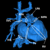
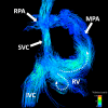
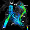
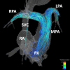
4D cardiovascular magnetic resonance velocity mapping of alterations of right heart flow patterns and main pulmonary artery hemodynamics in tetralogy of Fallot
- PMID: 22313680
- PMCID: PMC3305663
- DOI: 10.1186/1532-429X-14-16
4D cardiovascular magnetic resonance velocity mapping of alterations of right heart flow patterns and main pulmonary artery hemodynamics in tetralogy of Fallot
Abstract
Background: To assess changes in right heart flow and pulmonary artery hemodynamics in patients with repaired Tetralogy of Fallot (rTOF) we used whole heart, four dimensional (4D) velocity mapping (VM) cardiovascular magnetic resonance (CMR).
Methods: CMR studies were performed in 11 subjects with rTOF (5M/6F; 20.1 ± 12.4 years) and 10 normal volunteers (6M/4F; 34.2 ± 13.4 years) on clinical 1.5T and 3.0T MR scanners. 4D VM-CMR was performed using PC VIPR (Phase Contrast Vastly undersampled Isotropic Projection Reconstruction). Interactive streamline and particle trace visualizations of the superior and inferior vena cava (IVC and SVC, respectively), right atrium (RA), right ventricle (RV), and pulmonary artery (PA) were generated and reviewed by three experienced readers. Main PA net flow, retrograde flow, peak flow, time-to-peak flow, peak acceleration, resistance index and mean wall shear stress were quantified. Differences in flow patterns between the two groups were tested using Fisher's exact test. Differences in quantitative parameters were analyzed with the Kruskal-Wallis rank sum test.
Results: 4D VM-CMR was successfully performed in all volunteers and subjects with TOF. Right heart flow patterns in rTOF subjects were characterized by (a) greater SVC/IVC flow during diastole than systole, (b) increased vortical flow patterns in the RA and in the RV during diastole, and (c) increased helical or vortical flow features in the PA's. Differences in main PA retrograde flow, resistance index, peak flow, time-to-peak flow, peak acceleration and mean wall shear stress were statistically significant.
Conclusions: Whole heart 4D VM-CMR with PC VIPR enables detection of both normal and abnormal right heart flow patterns, which may allow for comprehensive studies to evaluate interdependencies of post-surgically altered geometries and hemodynamics.
Figures


References
-
- Lloyd-Jones D, Adams RJ, Brown TM, Carnethon M, Dai S, De Simone G, Ferguson TB, Ford E, Furie K, Gillespie C. et al. Heart disease and stroke statistics--2010 update: a report from the American Heart Association. Circulation. 2010;121:e46–e215. - PubMed
-
- Boechat MI, Ratib O, Williams PL, Gomes AS, Child JS, Allada V. Cardiac MR imaging and MR angiography for assessment of complex tetralogy of Fallot and pulmonary atresia. Radiographics. 2005;25:1535–1546. - PubMed
-
- Chowdhury UK, Pradeep KK, Patel CD, Singh R, Kumar AS, Airan B, Gulati GS, Kothari SS, Saxena A, Kalaivani M, Venugopal P. Noninvasive assessment of repaired tetralogy of Fallot by magnetic resonance imaging and dynamic radionuclide studies. Ann Thorac Surg. 2006;81:1436–1442. - PubMed
-
- Geva T, Sandweiss BM, Gauvreau K, Lock JE, Powell AJ. Factors associated with impaired clinical status in long-term survivors of tetralogy of Fallot repair evaluated by magnetic resonance imaging. J Am Coll Cardiol. 2004;43:1068–1074. - PubMed
-
- Oosterhof T, Mulder BJ, Vliegen HW, de Roos A. Cardiovascular magnetic resonance in the follow-up of patients with corrected tetralogy of Fallot: a review. Am Heart J. 2006;151:265–272. - PubMed
Publication types
MeSH terms
Substances
Grants and funding
LinkOut - more resources
Full Text Sources
Other Literature Sources
Medical

