. 2012 Feb 7;2(1):3.
doi: 10.1186/2044-5040-2-3.

TAK-1/p38/nNFκB signaling inhibits myoblast differentiation by increasing levels of Activin A

Affiliations

TAK-1/p38/nNFκB signaling inhibits myoblast differentiation by increasing levels of Activin A

Anne Ulrike Trendelenburg et al. Skelet Muscle. .

Abstract

Background: Skeletal-muscle differentiation is required for the regeneration of myofibers after injury. The differentiation capacity of satellite cells is impaired in settings of old age, which is at least one factor in the onset of sarcopenia, the age-related loss of skeletal-muscle mass and major cause of frailty. One important cause of impaired regeneration is increased levels of transforming growth factor (TGF)-β accompanied by reduced Notch signaling. Pro-inflammatory cytokines are also upregulated in aging, which led us hypothesize that they might potentially contribute to impaired regeneration in sarcopenia. Thus, in this study, we further analyzed the muscle differentiation-inhibition pathway mediated by pro-inflammatory cytokines in human skeletal muscle cells (HuSKMCs).

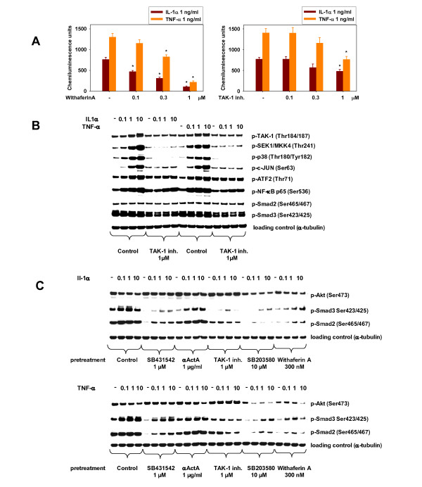
Methods: We studied the modulation of HuSKMC differentiation by the pro-inflammatory cytokines interleukin (IL)-1α and tumor necrosis factor (TNF)-α The grade of differentiation was determined by either imaging (fusion index) or creatine kinase (CK) activity, a marker of muscle differentiation. Secretion of TGF-β proteins during differentiation was assessed by using a TGF-β-responsive reporter-gene assay and further identified by means of pharmacological and genetic inhibitors. In addition, signaling events were monitored by western blotting and reverse transcription PCR, both in HuSKMC cultures and in samples from a rat sarcopenia study.

Results: The pro-inflammatory cytokines IL-1α and TNF-α block differentiation of human myoblasts into myotubes. This anti-differentiation effect requires activation of TGF-β-activated kinase (TAK)-1. Using pharmacological and genetic inhibitors, the TAK-1 pathway could be traced to p38 and NFκB. Surprisingly, the anti-differentiation effect of the cytokines required the transcriptional upregulation of Activin A, which in turn acted through its established signaling pathway: ActRII/ALK/SMAD. Inhibition of Activin A signaling was able to rescue human myoblasts treated with IL-1β or TNF-α, resulting in normal differentiation into myotubes. Studies in aged rats as a model of sarcopenia confirmed that this pro-inflammatory cytokine pathway identified is activated during aging.

Conclusions: In this study, we found an unexpected connection between cytokine and Activin signaling, revealing a new mechanism by which cytokines affect skeletal muscle, and establishing the physiologic relevance of this pathway in the impaired regeneration seen in sarcopenia.


Figures

Figure 1
Figure 1
Insulin-like growth factor (IGF)-1 and ALK inhibition counteracts interleukin (IL)-1α- and tumor necrosis factor (TNF)-α-induced inhibition of human skeletal muscle cell (HuSKMC) differentiation. (A) HuSKMC myotubes differentiated for 5 days in the absence (Con) and presence of IL-1α (0.1 ng/ml) and TNF-α (0.1 ng/ml), alone and in combination with IGF-1 (100 nmol/l) or SB431542 (1 μmol/l), were stained with anti-myosin heavy chain (MyHC) and DAPI. Shown are representative pictures and analysis of fusion index which was determined as the percentage of nuclei occurring in myotubes stained with MyHC on four pictures. Data are means ± SEM from three to six independent experiments. Differences from untreated HuSKMCs (control; first column),*P < 0.05; differences from IL-1α- and TNF-α-treated HuSKMCs (control; second and third column), #P < 0.05. (B) Analysis of creatine kinase (CK) activity in HuSKMC myotubes differentiated for 4 days and treated with either IL-1α (0.01-0.1 ng/ml) and TNF-α, alone (0.01 to 0.1 ng/ml) or in combination with IGF-1 (100 nmol/l) or SB431542 (1 μmol/l). Data are expressed as percentage of control untreated HuSKMCs. Data are means ± SEM from four to nine independent experiments. Differences from untreated HuSKMCs (control; first column),*P < 0.05; differences from IL-1α- and TNF-α-treated HuSKMCs (control; second to fourth columns), #P < 0.05.
Figure 2
Figure 2
Interleukin (IL)-1α and tumor necrosis factor (TNF)-α induced secretion of Activin A via activation of transforming growth factor-β-activated kinase (TAK)-1, p38 and nuclear factor (NF)κB pathways during human skeletal muscle cell (HuSKMC) differentiation. Supernatants (SNs) from HuSKMC myotubes differentiated for 3 days were analyzed in (A,B,D) HEK293Tcells stably transfected with the SMAD-sensitive CAGA-luc reporter in a reporter-gene assay (RGA) to assess occurrence of active TGF-β proteins or (C) with Activin A ELISA. (E) Activin A β-chain mRNA in HuSKMC myotubes differentiated for 6 hours was analyzed by reverse transcriptase (RT)-PCR. HuSKMCs were differentiated in the absence or presence of IL-1α (0.1 ng/ml) or TNF-α (0.1 ng/ml), alone or in combination (A-C) with the neutralizing anti-Activin A antibody (αActA), TAK-1 inhibitor, the p38 inhibitor SB203580 or the NFκB inhibitor withaferin A, or (D) after small interfering (si)RNA treatment. Data are expressed as (A,B,D) relative light units (RLU) or as percentage of control untreated HuSKMCs for (C) Activin A ELISA and (E) RT-PCR. Data are means ± SEM from 3 to 17 independent experiments. Differences from untreated HuSKMCs,*P < 0.05; differences from IL-1α- and TNF-α-treated HuSKMCs, #P < 0.05. (A) SNs were analyzed in CAGA-luc RGA. The soluble TGF-βRIIb/Fc chimera (500 ng/ml; TGF-βRIIb) was added alone to the supernatant to determine the contribution of the TGF-β isoforms (TGF-β1, TGF-β2 and TGF-β3) to CAGA-luc activity or in combination with αActA (10 μg/ml; TGF-βRIIb + αActA) to assess contribution of Activin A. (B) SNs were analyzed in CAGA-luc RGA. To eliminate responses to the TGF-β isoforms, CAGA-luc activity was measured after addition of TGF-βRIIb to the SN. (C) SNs were analyzed with Acitivin A ELISA. (D) SNs were analyzed in CAGA-luc RGA after addition of TGFβRIIb. SN were taken after treatment with siRNA against non-targeting control (siNTC), Activin A β-chain (siActivin A β-chain) or SMAD2 and SMAD3 (siSMAD2/3). (E) Activin A β-chain mRNA were analyzed by RT-PCR. Inhibitors were given 3 hours before IL-1α or TNF-α stimulation.
Figure 3
Figure 3
Inhibition of human skeletal muscle cell (HuSKMC) differentiation by interleukin (IL)-1α and tumor necrosis factor (TNF)-α is mediated by transforming growth factor-β-activated kinase (TAK)-1/p38/nuclear factor (NF)κB/Activin A/SMAD2/3 pathway. (A) HuSKMC myotubes differentiated for 5 days in the absence (Con) and presence of IL-1α (0.1 ng/ml) or TNF-α (0.1 ng/ml), alone and in combination with the neutralizing neutralizing anti-Activin A antibody (10 μg/ml; αActA) or TAK-1 inhibitor (1 μmol/l) were stained with anti-myosin heavy chain (MyHC) and DAPI. Shown are representative pictures and analysis of fusion index, which was determined as the percentage of nuclei occurring in myotubes stained with MyHC on four pictures taken. Data are means ± SEM from four to six independent experiments. Differences from untreated HuSKMCs (control; first column),*P < 0.05; differences from IL-1α- and TNF-α-treated HuSKMCs (control, second and third column), #P < 0.05. (B) Analysis of CK activity from in HuSKMC myotubes differentiated for 4 days and treated with either IL-1α (0.01-0.1 ng/ml) and TNF-α alone (0.01-0.1 ng/ml), and in combination with αActA (10 μg/ml), TAK-1 inhibitor (1 μmol/l), SB203580 (10 μmol/l) or withaferin A (100 nmol/l). Data are expressed as percentage of control from untreated HuSKMCs. Data are means ± SEM from three to nine independent experiments. Differences from untreated HuSKMCs (control; first column),*P < 0.05; differences from IL-1α- and TNF-α-treated HuSKMCs (control, second to fourth columns), #P < 0.05. (C) Analysis of CK activity from in HuSKMC myotubes differentiated for 4 days and treated with IL-1α (0.01-0.1 ng/ml) and TNF-α alone (0.01-0.1 ng/ml) after treatment with either small interfering (si)RNA against non-targeting control (siNTC), siActivin A β-chain or siSMAD2/3. Data are means ± SEM from five to seven independent experiments. Differences from siNTC-treated HuSKMCs (siNTC; first column),*P < 0.05; differences from IL-1α- and TNF-α-treated HuSKMCs (siNTC, second to fourth columns), #P < 0.05.
Figure 4
Figure 4
Inhibition of human skeletal muscle cell (HuSKMC) differentiation by interleukin (IL)-1α and tumor necrosis factor (TNF)-α is mediated by the transforming growth factor-β-activated kinase (TAK)-1/p38/nuclear factor (NF)κB/Activin A/SMAD2/3 pathway. (A) Analysis of NFκB-luc assay (RGA) from HuSKMC myoblasts treated with IL-1α (1 ng/ml) and TNF-α (1 ng/ml) alone, and in combination with TAK-1 inhibitor (0.1-1 μmol/l) or withaferin A (0.1-1 μmol/l). Data are expressed as relative light units (RLU). Data are means ± SEM from four independent experiments. Differences from IL-1α- and TNF-α-treated HuSKMCs (first column),*P < 0.05. (B) Immunoblotting of phospho-TAK-1, phospho-SEK1/MKK4, phospho-p38MAPK, phospho-c-Jun, phospho-ATF2, phospho-NFκB, phospho-SMAD2 and phospho-SMAD3 of samples from HuSKMCs treated with TNF-α (0.1 to 10 ng/ml) or IL-1α (0.1 to 10 ng/ml) for 15 minutes alone, and in the presence of TAK-1 inhibitor (1 μmol/l) starting at the onset of differentiation. The TAK-1 inhibitor was given 3 hours before IL-1α or TNF-α stimulation. Shown are representative immunoblots. (C) Immunoblotting of phospho-SMAD2, phospho-SMAD3 and pAKT of samples from HuSKMCs treated with TNF-α (0.1 to 10 ng/ml) or IL-1α (0.1 to 10 ng/ml) for 24 hours, alone and in the presence of SB431542 (1 μmol/l), αActA (10 μg/ml), TAK-1 inhibitor (1 μmol/l), SB203580 (10 μmol/l) or withaferin A (300 nmol/l) starting at the onset of differentiation. Inhibitors were given 3 hours before IL-1α or TNF-α stimulation. Shown are representative immunoblots.
Figure 6
Figure 6
Pathway model for interleukin (IL)-1α and tumor necrosis factor (TNF)-α-induced inhibition of human skeletal muscle cell (HuSKMC) differentiation. IL-1α and TNF-α induced secretion of Activin A in differentiating HuSKMCs cells and this secreted Activin A mediated their effects. Release of Activin A was dependent on transforming growth factor-β-activated kinase-1/p38/nuclear factor κB pathway activation, but independent of SMAD2/3 signaling. The released Activin A subsequently inhibited differentiation via ALK/SMAD2/3 signaling.
Figure 5
Figure 5
Transforming growth factor-β-activated kinase (TAK)-1/p38/Activin A/SMAD3 pathway activation in a rat sarcopenia model. (A) Immunoblotting of phospho-SMAD3, phospho-TAK-1, phospho-p38 and GAPDH in samples from rats of different ages. The gastrocnemius muscle was sampled from four rats per age point (6, 18, 12 and 24 months). Shown are representative immunoblots and averaged densitometry data from the blots. (B) Analysis of Activin A β-chain mRNA taken from rat gastrocnemius muscle. Data are expressed as percentage of control (first column, 6-month-old rats) and shown as means ± SEM from four individual rats. Differences from control (first column, 6-month-old rats),*P < 0.05.

References

    1. Clow C, Jasmin BJ. Skeletal muscle-derived BDNF regulates satellite cell differentiation and muscle regeneration. Mol Biol Cell. 2010. E10-02-0154. - PMC - PubMed
    1. Joe AWB, Yi L, Natarajan A, Le Grand F, So L, Wang J, Rudnicki MA, Rossi FMV. Muscle injury activates resident fibro/adipogenic progenitors that facilitate myogenesis. Nat Cell Biol. 2010;12(2):153–163. doi: 10.1038/ncb2015. - DOI - PMC - PubMed
    1. Conboy IM, Conboy MJ, Wagers AJ, Girma ER, Weissman IL, Rando TA. Rejuvenation of aged progenitor cells by exposure to a young systemic environment. Nature. 2005;433(7027):760. doi: 10.1038/nature03260. - DOI - PubMed
    1. Coleman ME, DeMayo F, Yin KC, Lee HM, Geske R, Montgomery C, Schwartz RJ. Myogenic vector expression of insulin-like growth factor I stimulates muscle cell differentiation and myofiber hypertrophy in transgenic mice. J Biol Chem. 1995;270(20):12109–12116. doi: 10.1074/jbc.270.20.12109. - DOI - PubMed
    1. Florini JR, Ewton DZ, Coolican SA. Growth hormone and the insulin-like growth factor system in myogenesis. Endocr Rev. 1996;17(5):481–517. - PubMed