. 2012 Apr 24;26(7):805-14.
doi: 10.1097/QAD.0b013e328351f780.

A role for cytomegalovirus-specific CD4+CX3CR1+ T cells and cytomegalovirus-induced T-cell immunopathology in HIV-associated atherosclerosis

Affiliations

A role for cytomegalovirus-specific CD4+CX3CR1+ T cells and cytomegalovirus-induced T-cell immunopathology in HIV-associated atherosclerosis

Karim Sacre et al. AIDS. .

Abstract

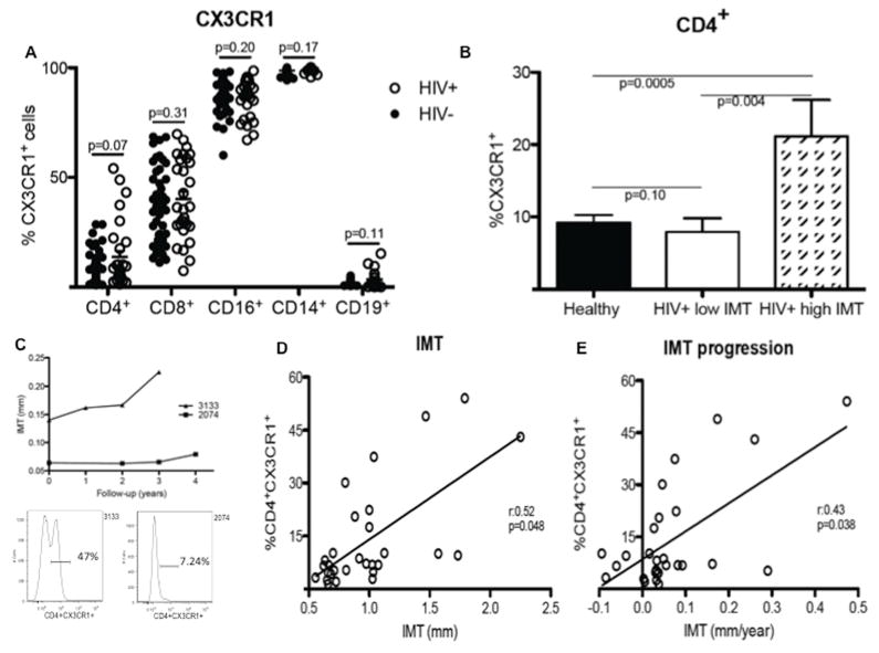
Objective: HIV-infected individuals are at increased risk for myocardial infarction. Given observations that cytomegalovirus (CMV) infection, CMV-specific T cells, and CX3CR1 have each been associated with atherosclerosis, we hypothesized that CMV-induced T-cell immunopathology could contribute to HIV-associated atherosclerosis.

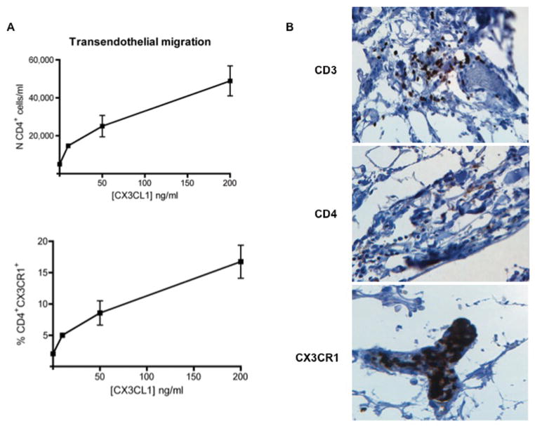
Methods: We measured the expression of CX3CR1 on peripheral blood mononuclear cells and its association with carotid artery intima-media thickness (IMT) in 29 HIV-infected individuals and 48 uninfected controls. We analyzed the phenotype and specificity of CX3CR1(+)CD4(+) T cells, the production of CX3CL1 (the ligand of CX3CR1) by CMV-infected endothelial cells in vitro, and the migration of CD4(+) T cells induced by CX3CL1.

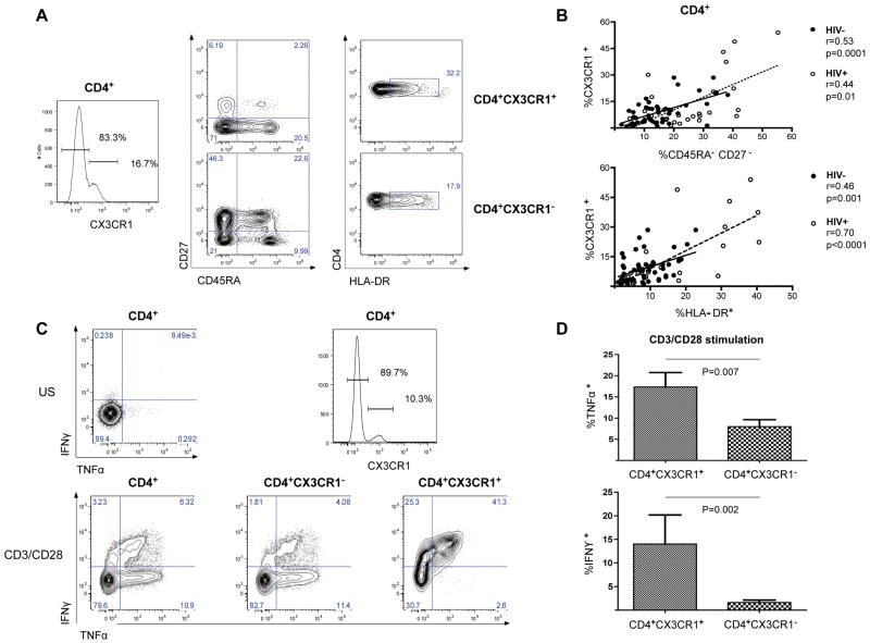
Results: The progression of atherosclerosis in HIV-infected individuals, as assessed by longitudinal measurements of carotid IMT, was associated with a high frequency of CD4(+) T cells that express the chemokine receptor CX3CR1. Such CD4(+)CX3CR1(+) T cells were antigen-primed, produced high levels of pro-inflammatory cytokines, and composed the majority of the CMV-specific CD4(+) T cells. CMV-stimulated CD4(+) T cells were also found to induce the production of CX3CL1 (the ligand for CX3CR1) by human arterial endothelial cells, driving the transendothelial migration of pro-inflammatory CD4(+) T cells. Finally, we observed that CD4(+)CX3CR1(+) T cells could be localized to the coronary arterial wall in HIV disease.

Conclusion: HIV-associated atherosclerosis may be driven by a positive feedback pathway in which a high frequency of antigen-stimulated, CMV-specific CD4(+)CX3CR1(+) T cells induce endothelial cells to secrete CX3CL1, which itself drives progressive infiltration of the arterial wall by pro-inflammatory cells.


Conflict of interest statement

There are no financial conflicts of interest associated with this work.

Figures

Figure 1
Figure 1. High percentage of CD4+CX3CR1+ T cells in HIV-infected subjects correlates with IMT progression
(A) Flow cytometric analysis of CX3CR1 expression on CD4+, CD8+, CD16+, CD14+, and CD19+ cells found in PBMCs of HIV-uninfected (HIV−, n=48, blacks circles) and HIV-infected (HIV+, n=29, white circles) subjects. (B) Comparison of the frequency of CX3CR1+CD4+ T cells in HIV and HIV+ subjects separated into two groups: one with low (<1mm) and another with high (≥1mm) IMT. (C) Carotid artery IMT progression over time in two HIV+ subjects, one (3133) showing a high frequency (47%; see flow cytogram on left) of CX3CR1+CD4+ T cells and the other (2074) showing a low frequency (7.24%; see flow cytogram on right) of CD4+CX3CR1+ T cells. (D) Positive statistical correlation between the frequency of CX3CR1+CD4+ T cells and IMT measurement at baseline. (E) IMT progression over time in HIV+ subjects. Significant correlations were found after adjustment for age.
Figure 2
Figure 2. Phenotype and functional characteristics of CD4+CX3CR1+ T cells
(A) Flow cytometric analysis of CD27 and CD45RA (middle) and of HLA-DR (right) co-expression on resting CD4+CX3CR1+ (top) and CD4+CX3CR1 T cells (bottom) from a representative subject. (B) Positive statistical correlation in both groups (HIV− and HIV+ subjects) between the expression of CX3CR1 and the lack of expression of CD27 and CD45RA (top) and the expression of HLA-DR (bottom) on CD4+ T cells. (C) Flow cytometric analysis of TNFα and IFNγ production after TCR stimulation (using CD3/CD28 stimulation) of CD4+ T cells (left), CD4+CX3CR1 T cells (middle), and CD4+CX3CR1+ T cells (right) from a representative subject. CX3CR1 expression was defined as per the histogram on the upper right, with an unstimulated (US) control in the panel to the upper left. (D) Mean production of TNFα (top) and IFNγ (bottom) by CD4+CX3CR1+ T cells and CD4+CX3CR1 T cells upon polyclonal stimulation, as analyzed in 12 subjects.
Figure 3
Figure 3. CD4+CX3CR1+ T cells are CMV-specific and CX3CL1 production is induced in CMV-infected human artery endothelial cells
(A) Flow cytometric analysis of TNFα and IFNγ production after CMV-pp65 stimulation of CD4+CX3CR1 T cells (left panel), and CD4+CX3CR1+ T cells (right panel) from a representative subject. CX3CR1 expression was defined as per the histogram above the two panels. (B) Mean production of TNFα (left) and IFNγ (right) by CD4+CX3CR1+ T cells and CD4+CX3CR1 T cells upon CMV-pp65 stimulation, as analyzed in 16 HIV-infected subjects. (C) Secretion of CX3CL1 (as detected by ELISA) into the supernatant of uninfected HAEC (HAECCMV−), CMV-infected HAEC (HAECCMV+), uninfected HAEC co-cultured with PBMCs from CMV-positive subjects (HAECCMV− PBMCs), and CMV-infected HAEC co-cultured with PBMCs from CMV-positive subjects (HAECCMV+ PBMCs). (D) Secretion of CX3CL1 (as detected by ELISA) into the supernatant of HAEC cultured in the same conditions as described above and in the supernatant of CMV-infected HAEC (HAECCMV+) co-cultured with either CD4-depleted PBMCs (CD4) or with PBMCs and neutralizing antibodies against TNFα (anti-TNFα) or IFNγ (anti-IFNγ), or with isotype control antibodies (IgG). Neutralizations were carried out directly in co-cultivations of PBMCs with HAECCMV+ at a concentration of 30 μg/ml (for anti-TNFα) or 20 μg/ml (for anti-IFNγ). The data are representative of four separate experiments with different blood donors and CMV-infected HAEC monolayers. *, means p<0.05; **, means p>0.005; ns, means not significant
Figure 4
Figure 4. CX3CL1 supports chemoattraction of CD4+ T cells
(A) Transmigrated CD4+ T cells from transendothelial migration assays incubated with varying concentration of CX3CL1 (10 ng/ml, 50 ng/ml, and 200 ng/ml) and with control media, expressed as the absolute number of CD4+ T cells/ml (top) or as the percentage of input CD4+ T cells expressing CX3CR1 (bottom). For each sample, three transmigration wells were set up and the transmigrated populations were counted from each well independently. Results are representative of three independent assays. (B) Immunohistochemical detection of CD3, CD4, and CX3CR1 in adjacent sections of the same coronary artery showing early atheromatous lesions from an HIV+ subject. An aggregate of CD3+ immunoreactive cells is observed in the perivascular space of a small blood vessel running in the adventitia of the artery (×20, top). CD4+ cells (x20, middle) and CX3CR1+ cells (x20, bottom) are scattered in the wall and present in the lumen of a small blood vessel running in the adventitia of the artery, respectively.

References

    1. Hsue PY, Lo JC, Franklin A, Bolger AF, Martin JN, Deeks SG, et al. Progression of atherosclerosis as assessed by carotid intima-media thickness in patients with HIV infection. Circulation. 2004;109:1603–1608. - PubMed
    1. Barbaro G, Fisher SD, Lipshultz SE. Pathogenesis of HIV-associated cardiovascular complications. Lancet Infect Dis. 2001;1:115–124. - PubMed
    1. Durand M, Sheehy O, Baril JG, Lelorier J, Tremblay CL. Association between HIV infection, antiretroviral therapy, and risk of acute myocardial infarction: a cohort and nested case-control study using Quebec’s public health insurance database. Journal of Acquired Immune Deficiency Syndromes. 2011;57:245–253. - PubMed
    1. Baker JV, Henry WK, Patel P, Bush TJ, Conley LJ, Mack WJ, et al. Progression of carotid intima-media thickness in a contemporary human immunodeficiency virus cohort. Clinical infectious diseases : an official publication of the Infectious Diseases Society of America. 2011;53:826–835. - PMC - PubMed
    1. van Vonderen MG, Hassink EA, van Agtmael MA, Stehouwer CD, Danner SA, Reiss P, et al. Increase in carotid artery intima-media thickness and arterial stiffness but improvement in several markers of endothelial function after initiation of antiretroviral therapy. The Journal of Infectious Diseases. 2009;199:1186–1194. - PubMed

Publication types

MeSH terms