. 2012 Feb 7;14(1):R23.
doi: 10.1186/bcr3108.

Contribution of CXCL12 secretion to invasion of breast cancer cells

Affiliations

Contribution of CXCL12 secretion to invasion of breast cancer cells

Pamela J Boimel et al. Breast Cancer Res. .

Abstract

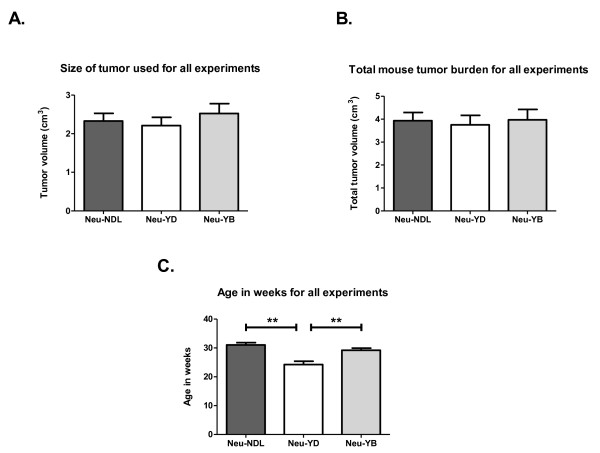
Introduction: Neu (HER2/ErbB2) is overexpressed in 25% to 30% of human breast cancer, correlating with a poor prognosis. Researchers in previous studies who used the mouse mammary tumor virus Neu-transgenic mouse model (MMTV-Neu) demonstrated that the Neu-YB line had increased production of CXCL12 and increased metastasis, whereas the Neu-YD line had decreased metastasis. In this study, we examined the role of increased production of CXCL12 in tumor cell invasion and malignancy.

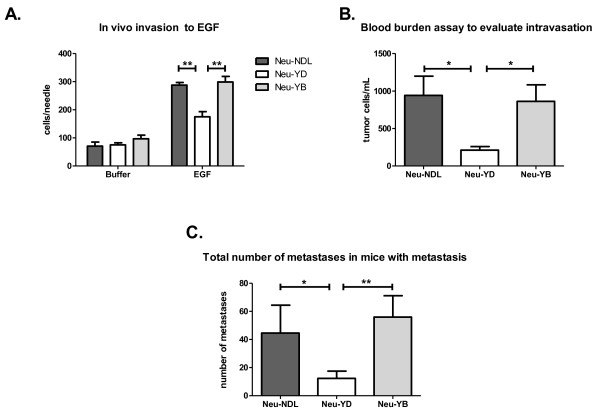
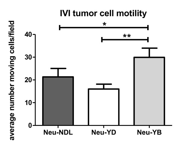
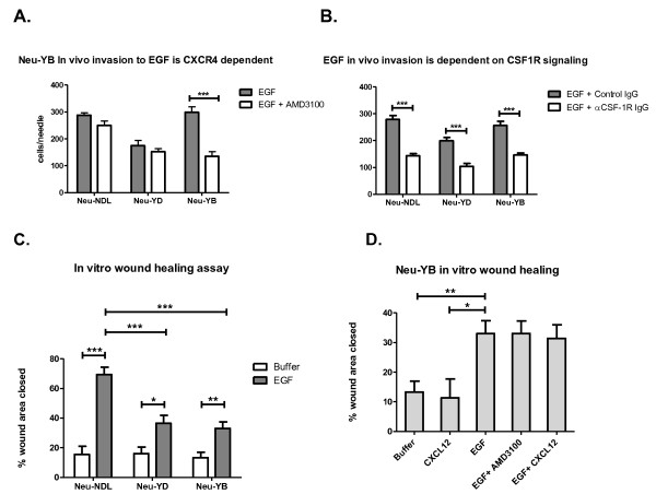
Methods: We studied invasion in the tumor microenvironment using multiphoton intravital imaging, in vivo invasion and intravasation assays. CXCL12 signaling was altered by using the CXCR4 inhibitor AMD3100 or by increasing CXCL12 expression. The role of macrophage signaling in vivo was determined using a colony-stimulating factor 1 receptor (CSF-1R) blocking antibody.

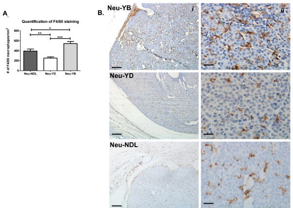
Results: The Neu-YD strain was reduced in invasion, intravasation and metastasis compared to the Neu-YB and Neu deletion mutant (activated receptor) strains. Remarkably, in the Neu-YB strain, in vivo invasion to epidermal growth factor was dependent on both CXCL12-CXCR4 and CSF1-CSF-1R signaling. Neu-YB tumors had increased macrophage and microvessel density. Overexpression of CXCL12 in rat mammary adenocarcinoma cells increased in vivo invasion as well as microvessel and macrophage density.

Conclusions: Expression of CXCL12 by tumor cells results in increased macrophage and microvessel density and in vivo invasiveness.


Figures

Figure 1
Figure 1
Evaluation of tumor growth, burden and age. (A) Size of largest tumor at time of analysis. There was no significant difference between the Neu deletion mutant (activated receptor) (Neu-NDL), Neu-YD and Neu-YB strains. (B) Total tumor volume at time of analysis. Transgenic mice developed multiple tumors. All tumors were measured, and the volumes were added. There was no significant difference in cumulative tumor volume, which averaged about 4 ml. (C) Average age in weeks at time of analysis. Data are means and SEM. **P < 0.005 by t-test, n = 18 to 22 mice per strain.
Figure 2
Figure 2
Neu-YD strain exhibits decreased invasion, intravasation and metastasis compared to Neu-NDL and Neu-YB strains. (A) The in vivo invasion assay was performed, allowing tumor cells to invade over 4 hours into microneedles containing 25 nM epidermal growth factor (EGF) or buffer. Neu-NDL = Neu deletion mutant (activated receptor). Data are means and SEM. **P < 0.005 by t-test, n = three to five mice per strain and five to seven needles per condition. (B) A blood burden assay was performed to evaluate intravasation. The number of single cells per milliliter of blood were counted. Data are means and SEM. *P < 0.05 by t-test, n = 8 to 11 mice per strain. (C) H & E-stained sections (5 μm each) spaced 250 μm apart were taken from the whole lung sample, and metastases were counted in every section. The efficiency of lung metastasis is represented as the total number of metastases in all lung sections for each animal that had metastases. Data are means and SEM. *P < 0.05 and ** p < 0.005 by Mann-Whitney U test; n = 16 Neu-NDL mice, 15 Neu-YD mice and 8 Neu-YB mice.
Figure 3
Figure 3
Motility in the tumor microenvironment. Intravital imaging (IVI) using multiphoton microscopy of primary mammary tumors from transgenic Neu deletion mutant (activated receptor) (Neu-NDL), Neu-YD and Neu-YB cyan fluorescent protein mice. Thirty-minute time-lapse Z-series were collected, and the average total cell motility was quantified per 50-μm Z-stack (five sections imaged at 10-μm intervals). Data are means and SEM of 27 to 32 separate Z-stacks. *P < 0.05, **P < 0.005 by Mann-Whitney U test; n = 13 Neu-NDL mice, n = 8 Neu-YD mice and n = 12 Neu-YB mice.
Figure 4
Figure 4
F4/80 staining indicates the Neu-YB strain recruits more macrophages into the tumor parenchyma. (A) F4/80 staining was quantified by using a 40× lens objective and counting the number of F4/80-stained macrophages per field (n = 3 tumors per strain and 10 random fields per tumor). Data shown are means and SEM. *P < 0.05, **P < 0.005, ***P < 0.0005 by t-test. (B) Representative fields of F4/80 staining taken at low magnification (scale bar = 200 μm) (i) and high magnification (scale bar = 25 μm) (ii). Neu-NDL = Neu deletion mutant (activated receptor).
Figure 5
Figure 5
Epidermal growth factor-induced in vivo invasion is dependent on CXCR4/CXCL12 paracrine signaling in the tumor microenvironment. (A) In vivo invasion assay performed by infusing 25 nM epidermal growth factor (EGF) and EGF + 0.5 μM CXCR4 inhibitor AMD3100 through microneedles inserted into the primary tumor to collect the invasive cell population over 4 hours. Neu-NDL = Neu deletion mutant (activated receptor). Data are means and SEM. ***P < 0.0005 by t-test, n = three to five mice per strain and five to seven needles per condition. (B) In vivo invasion assay performed with 25 nM EGF + control rat immunoglobulin G (IgG) antibody or EGF + colony-stimulating factor 1 receptor (CSF-1R) blocking antibody (αCSF-1R IgG) in the needles. Data are means and SEM. ***P < 0.0005 by t-test, n = three to five mice per strain and five to seven needles per condition. (C) In vitro wound healing assays in the absence (Buffer) or presence of 5 nM EGF (EGF). Data are means and SEM. *P < 0.05, **P < 0.005 and ***P < 0.0005 by t-test, n = 3 tumors per strain and at least 10 fields per condition. (D) In vitro wound healing assays were performed with confluent primary tumor tissue in the presence of buffer (Buffer), 5 nM EGF alone (EGF), 5 nM EGF with 100 nM AMD3100 (EGF + AMD3100), 5 nM EGF with 1 nM CXCL12 (EGF + CXCL12) or 1 nM CXCL12 alone (CXCL12). Data are means and SEM. **P < 0.005 by t-test, n = 3 tumors and at least 10 fields per condition.
Figure 6
Figure 6
Rat mammary adenocarcinoma CXCL12 cells show increased invasion to epidermal growth factor and recruitment of macrophages. (A) The in vivo invasion assay was performed with MTLn3 empty vector and CXCL12-overexpressing cell line tumors, allowing tumor cells to invade over 4 hours into microneedles containing 25 nM EGF or buffer. Data shown are means and SEM. *P < 0.05 and ***P < 0.0005 by t-test. (B) F4/80 staining was quantified by using a 40× lens objective and counting the number of F4/80-stained macrophages per field (n = 5 tumors per cell line and 10 random fields per tumor). Data shown are means and SEM. **P < 0.005 by t-test. (C) Representative images of F4/80 staining for macrophages in MTLn3 pQCXIP and MTLn3 pQCXIP-CXCL12 tumors. Images were taken using a 40× lens objective. Scale bar = 25 μm.

References

    1. Slamon DJ, Godolphin W, Jones LA, Holt JA, Wong SG, Keith DE, Levin WJ, Stuart SG, Udove J, Ullrich A, Press MF. Studies of the HER-2/neu proto-oncogene in human breast and ovarian cancer. Science. 1989;244:707–712. doi: 10.1126/science.2470152. - DOI - PubMed
    1. Yarden Y. The EGFR family and its ligands in human cancer: signalling mechanisms and therapeutic opportunities. Eur J Cancer. 2001;37(Suppl 4):S3–S8. - PubMed
    1. Klapper LN, Glathe S, Vaisman N, Hynes NE, Andrews GC, Sela M, Yarden Y. The ErbB-2/HER2 oncoprotein of human carcinomas may function solely as a shared coreceptor for multiple stroma-derived growth factors. Proc Natl Acad Sci USA. 1999;96:4995–5000. doi: 10.1073/pnas.96.9.4995. - DOI - PMC - PubMed
    1. Tzahar E, Waterman H, Chen X, Levkowitz G, Karunagaran D, Lavi S, Ratzkin BJ, Yarden Y. A hierarchical network of interreceptor interactions determines signal transduction by Neu differentiation factor/neuregulin and epidermal growth factor. Mol Cell Biol. 1996;16:5276–5287. - PMC - PubMed
    1. Pinkas-Kramarski R, Soussan L, Waterman H, Levkowitz G, Alroy I, Klapper L, Lavi S, Seger R, Ratzkin BJ, Sela M, Yarden Y. Diversification of Neu differentiation factor and epidermal growth factor signaling by combinatorial receptor interactions. EMBO J. 1996;15:2452–2467. - PMC - PubMed

Publication types

MeSH terms