. 2012 Apr 4;31(7):1714-26.
doi: 10.1038/emboj.2012.19. Epub 2012 Feb 7.

MDA5 cooperatively forms dimers and ATP-sensitive filaments upon binding double-stranded RNA

Affiliations

MDA5 cooperatively forms dimers and ATP-sensitive filaments upon binding double-stranded RNA

Ian C Berke et al. EMBO J. .

Abstract

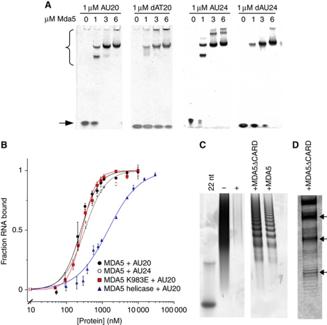
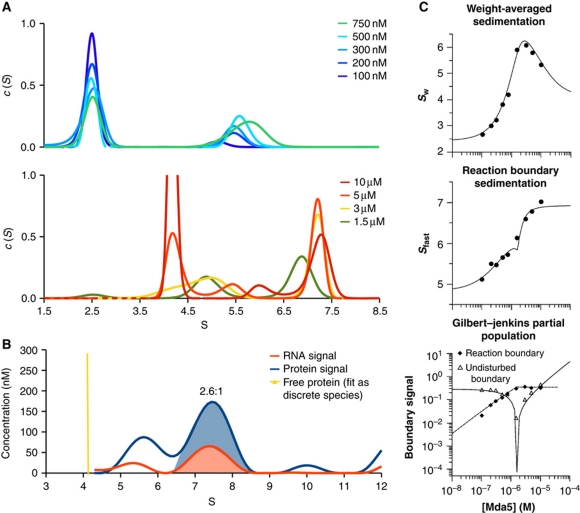
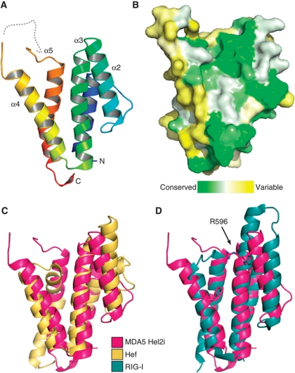
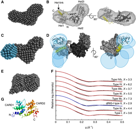
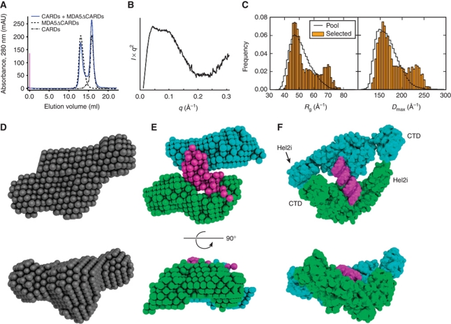
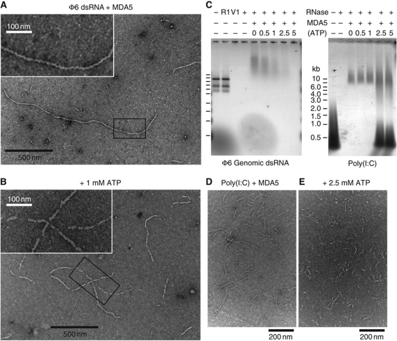
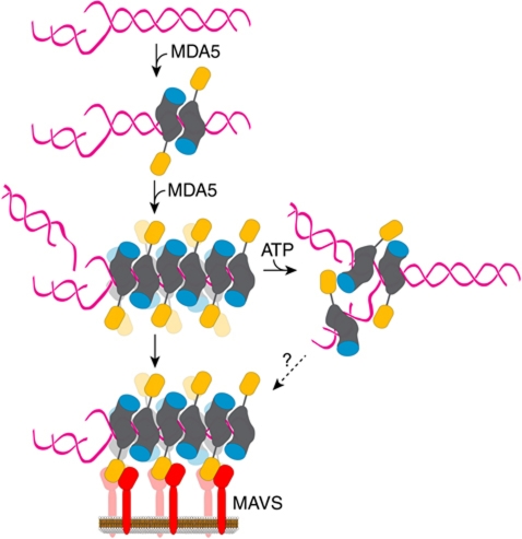
Melanoma differentiation-associated gene-5 (MDA5) detects viral double-stranded RNA in the cytoplasm. RNA binding induces MDA5 to activate the signalling adaptor MAVS through interactions between the caspase recruitment domains (CARDs) of the two proteins. The molecular mechanism of MDA5 signalling is not well understood. Here, we show that MDA5 cooperatively binds short RNA ligands as a dimer with a 16-18-basepair footprint. A crystal structure of the MDA5 helicase-insert domain demonstrates an evolutionary relationship with the archaeal Hef helicases. In X-ray solution structures, the CARDs in unliganded MDA5 are flexible, and RNA binds on one side of an asymmetric MDA5 dimer, bridging the two subunits. On longer RNA, full-length and CARD-deleted MDA5 constructs assemble into ATP-sensitive filaments. We propose a signalling model in which the CARDs on MDA5-RNA filaments nucleate the assembly of MAVS filaments with the same polymeric geometry.


Conflict of interest statement

The authors declare that they have no conflict of interest.

Figures

Figure 1
Figure 1
MDA5 binds short nucleic acid ligands with a 16–18-bp footprint. (A) Native PAGE showing the mobility shift of 20- and 24-bp RNA and DNA oligonucleotides (stained with SYBR Green) by full-length MDA5. MDA5–oligonucleotide complexes (bracket) migrate more slowly than free oligonucleotides (arrow). (B) Binding curves based on densitometry of bands in EMSA. MDA5 bound AU20 with Kd=287±13 nM and a Hill coefficient, n=1.7 (black squares). MDA5 bound AU24 with Kd=360±22 nM, n=1.5 (open squares). MDA5 K983E bound AU20 with Kd=277±14.8 nM, n=2.1 (red). The MDA5 helicase construct bound AU20 with Kd=1.45±0.05 μM, n=1 (blue). Each curve is the mean±s.e.m. of three to four experiments. (C) Poly(I:C) RNase protection with full-length and CARD-deleted MDA5 on denaturing PAGE. Both constructs protect poly(I:C) from RNases I and V1 in 16–18-bp increments with the smallest band at ∼50 nt. Poly(I:C) (− lane) is completely degraded by RNases (+ lane). Left lane, 22-bp DNA marker. (D) High-resolution gel showing the fine structure of bands in the RNase protection experiment from (C). See also Supplementary Figure S1.
Figure 2
Figure 2
Short dsRNA induces rapid and cooperative dimerization of CARD-deleted MDA5. (A) The sedimentation coefficient distributions, c(S), calculated from SV-AUC with 1 μM AU20 RNA monitored at 260 nm. Top and bottom panels, c(S) for MDA5 concentrations below and above 1 μM, respectively. Free AU20 sediments at 2.4 S and free MDA5 at 4.2 S. The limiting peak at 7.4 S (bottom panel) is consistent with a 2:1 MDA5:AU20 complex. (B) Multisignal SV-AUC with 15 μM MDA5 and 1 μM AU20 monitored at 260 nm and with interference optics indicates a 2.6:1 MDA5:AU20 stoichiometry within the complex, based on integration of the areas under the 7.4 S peaks. (C) Global isotherm analysis using the parameters from the curves in (A). The weight-averaged sedimentation coefficient (top), reaction boundary sedimentation coefficient (middle) and population signals of the undisturbed and reaction boundaries (bottom) yielded dissociation constants of 187 and 124 nM for association to AU20 of the first and second MDA5 subunits, respectively. Models enforcing completely independent binding sites (Kd2/Kd1=4) had significantly worse fits (Supplementary Figure S2).
Figure 3
Figure 3
Crystal structure of the MDA5 helicase-insert (Hel2i) domain. (A) Cartoon representation of MDA5 Hel2i (blue to red, N- to C-terminus). The disordered loop containing the ΔL2 loop deletion is shown as a dashed line. (B) Surface representation coloured according to sequence homology across vertebrate MDA5 sequences (green is conserved, yellow is variable). The view is the same as in (A). (C) MDA5 Hel2i (magenta) has the same overall topology as P. furiousus Hef Hel2i (yellow), with an RMSD of Cα atom positions of 3.8 Å. (D) Comparison of MDA5 Hel2i and duck RIG-I Hel2i (blue). Two residues important for binding of Hel2i to the CARDs in RIG-I (F540 and F571) are shown in stick representation with the corresponding residues for MDA5 (R596 and F630). The extended α3 helix in RIG-I also contributes to the CARD interface. See also Supplementary Table SI.
Figure 4
Figure 4
SAXS structures of unliganded MDA5 fragments. (A) DA model of the MDA5 helicase domains. (B) Homology model of the Hel1, Hel2i and Hel2 helicase domains (light to dark grey, respectively) refined against the SAXS curve. The helix between Hel2 and the CTD (Hel-link; yellow) was docked manually prior to refinement. The envelope of the DA model is shown superimposed. (C) DA model of CARD-deleted MDA5 with the additional lobe corresponding to the CTD in cyan. (D) Superposition of the six best rigid-body models of CARD-deleted MDA5 consistent with the SAXS data. Coloured regions were constrained by flexible loops; the helicase domains (grey) were fixed. The positions of the CTDs are indicated by ovals and a representative cartoon (cyan). (E) DA model of the CARDs. All DA models represent an average of 15 models. (F) Calculated one-dimensional X-ray scattering of MDA5 CARD homology models in prototypical death domain family interactions (red and blue) fitted against the observed scattering (black). The ‘b’ indicates a swap of the two CARD domains across the interface. The curves were vertically offset for clarity and the best fit is indicated in blue. (G) Cartoon representation of the CARD homology model with the type II interface (blue to red, N- to C-terminus). See also Supplementary Figure S3 and Supplementary Table SII.
Figure 5
Figure 5
The MDA5 helicase and CTD domains do not associate with the CARDs and bind RNA as a dimer. (A) The MDA5 CARDs and CARD-deleted MDA5 elute separately from a size-exclusion column (blue). Elution curves of the two components loaded separately are shown in grey. (B) Plateau in the Kratky plot of full-length MDA5 indicates substantial flexibility within the molecule. (C) Histograms of the radius of gyration (Rg) and maximum dimension (Dmax) of full-length MDA5 models (orange) selected to fit the SAXS data with a five-curve minimal ensemble from a pool of 105 random domain arrangements (black). (D) Single-phase DA model of CARD-deleted MDA5 in complex with AU20 using data to 0.168 Å−1 (average of 15 models). (E) Three-phase DA model (average of seven) of the same complex as in (D) with two protein phases (cyan and green) and one RNA phase (magenta). (F) Rigid-body model using knowledge-based restraints of the same complex as in (D, E). See Supplementary Figures S4 and S5.
Figure 6
Figure 6
MDA5 assembles into regular filaments on long dsRNA ligands. (A) Negative-stain electron micrograph (EM) of MDA5 mixed with bacteriophage ϕ6 genomic dsRNA shows filaments 8–10 nm in diameter. Filaments have few breaks along their length and can be up to 1.8 μm long. Inset: close-up of the boxed region. (B) 1 mM ATP induces breaks in the filaments. At 5 mM ATP, no filaments were observed. Inset: close-up of the boxed region. (C) Nuclease protection assay shows ϕ6 RNA is not protected by MDA5 when ATP is present (left). The three ϕ6 genome segments (2.9, 4.1 and 6.4 kb) are degraded by dsRNA endonuclease V1 (V1), but not ssRNA endonuclease 1 (R1). MDA5 reduces the mobility of ϕ6 and poly(I:C) RNA on agarose gel electrophoresis and protects them from RNases. Increasing ATP levels decrease RNase protection by MDA5 of ϕ6 RNA (left) but not of poly(I:C) RNA (right). At >2.5 mM ATP, free poly(I:C) is also protected. (D, E) EM of poly(I:C) and MDA5 without ATP (D), and with 2.5 mM ATP (E). Filaments incubated with ATP tend to aggregate, most likely from enhanced filament crosslinking. This effect is not observed with AMPPNP. See Supplementary Figure S6 for protein-only and RNA-only controls.
Figure 7
Figure 7
Proposed model for MDA5 ligand binding and activation. MDA5 binds dsRNA cooperatively as a dimer with a 16–18-bp footprint. The dimer contacts are formed by the helicase domains (grey) and the CTD (cyan). The antiparallel arrangement of the subunits within the dimers is based on the homology model of a CARD-deleted MDA5 dimer bound to RNA refined against SAXS data. The binding mode of full-length MDA5 may differ from that shown. Full-length (or CARD-deleted) MDA5 assembles into helical or linear polymeric filaments on long dsRNA ligands. The CARDs (orange) on the outside of filaments recruit the CARD of MAVS (red) on the mitochondrial membrane. Through this CARD–CARD interaction, the regular helical or linear arrangement of MDA5 CARDs is imposed on MAVS, thereby inducing the formation of MAVS fibrils, which propagate the signalling response (Hou et al, 2011).

References

    1. Aida K, Nishida Y, Tanaka S, Maruyama T, Shimada A, Awata T, Suzuki M, Shimura H, Takizawa S, Ichijo M, Akiyama D, Furuya F, Kawaguchi A, Kaneshige M, Itakura J, Fujii H, Endo T, Kobayashi T (2011) RIG-I- and MDA5-initiated innate immunity linked with adaptive immunity accelerates beta-cell death in fulminant type 1 diabetes. Diabetes 60: 884–889 - PMC - PubMed
    1. Bamming D, Horvath CM (2009) Regulation of signal transduction by enzymatically inactive antiviral RNA helicase proteins MDA5, RIG-I, and LGP2. J Biol Chem 284: 9700–9712 - PMC - PubMed
    1. Baum J, Tonkin CJ, Paul AS, Rug M, Smith BJ, Gould SB, Richard D, Pollard TD, Cowman AF (2008) A malaria parasite formin regulates actin polymerization and localizes to the parasite-erythrocyte moving junction during invasion. Cell Host Microbe 3: 188–198 - PubMed
    1. Binder M, Eberle F, Seitz S, Mucke N, Huber CM, Kiani N, Kaderali L, Lohmann V, Dalpke A, Bartenschlager R (2011) Molecular mechanism of signal perception and integration by the innate immune sensor retinoic acid inducible gene-I (RIG-I). J Biol Chem 286: 27278–27287 - PMC - PubMed
    1. Brown PH, Balbo A, Schuck P (2009) On the analysis of sedimentation velocity in the study of protein complexes. Eur Biophys J 38: 1079–1099 - PMC - PubMed

Publication types