. 2012 May 9;18(1):477-85.
doi: 10.2119/molmed.2012.00024.

Inhibition of high-mobility group box 1 protein (HMGB1) enhances bacterial clearance and protects against Pseudomonas Aeruginosa pneumonia in cystic fibrosis

Affiliations

Inhibition of high-mobility group box 1 protein (HMGB1) enhances bacterial clearance and protects against Pseudomonas Aeruginosa pneumonia in cystic fibrosis

Maria Entezari et al. Mol Med. .

Abstract

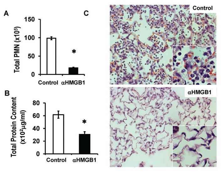
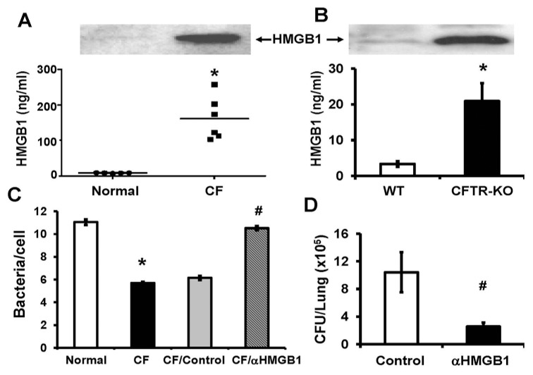
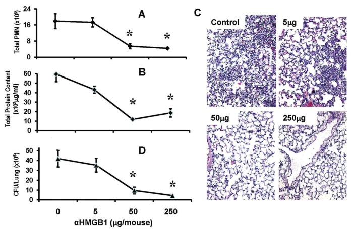
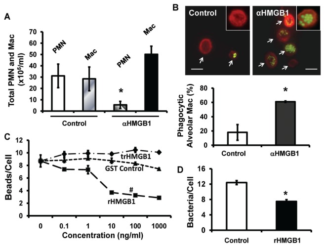
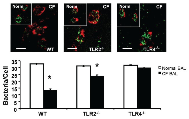
Pulmonary infection with Pseudomonas aeruginosa and neutrophilic lung inflammation significantly contribute to morbidity and mortality in cystic fibrosis (CF). High-mobility group box 1 protein (HMGB1), a ubiquitous DNA binding protein that promotes inflammatory tissue injury, is significantly elevated in CF sputum. However, its mechanistic and potential therapeutic implications in CF were previously unknown. We found that HMGB1 levels were significantly elevated in bronchoalveolar lavage fluids (BALs) of CF patients and cystic fibrosis transmembrane conductance regulator (CFTR )(-/-) mice. Neutralizing anti-HMGB1 monoclonal antibody (mAb) conferred significant protection against P. aeruginosa-induced neutrophil recruitment, lung injury and bacterial infection in both CFTR(-/-) and wild-type mice. Alveolar macrophages isolated from mice treated with anti-HMGB1 mAb had improved phagocytic activity, which was suppressed by direct exposure to HMGB1. In addition, BAL from CF patients significantly impaired macrophage phagocytotic function, and this impairment was attenuated by HMGB1-neutralizing antibodies. The HMGB1-mediated suppression of bacterial phagocytosis was attenuated in macrophages lacking toll-like receptor (TLR)-4, suggesting a critical role for TLR4 in signaling HMGB1-mediated macrophage dysfunction. These studies demonstrate that the elevated levels of HMGB1 in CF airways are critical for neutrophil recruitment and persistent presence of P. aeruginosa in the lung. Thus, HMGB1 may provide a therapeutic target for reducing bacterial infection and lung inflammation in CF.


Figures

Figure 1
Figure 1
Anti-HMGB1 mAb reduces P. aeruginosa–induced pulmonary neutrophil recruitment and lung injury in CFTR−/− mice. Male CFTR−/− mice were treated with 250 μg/mouse of either anti-HMGB1 mAb (αHMGB1) or isotype control antibody (control) 12 h before and at the time of inoculation with GFP-expressing P. aeruginosa PAO1. Eighteen hours later, mice were euthanized, and lungs and BALs were harvested. Neutrophilic inflammation was assessed by the PMN in the BAL (A). Total protein content in BAL (B) and histological analysis (C) were used to assess lung injury. Mean ± SEM, *P < 0.05 (n = 5–6 mice per group of three independent experiments). (C) Representative images of HE-staining lung sections.
Figure 2
Figure 2
Elevated levels of HMGB1 in BAL of CF patients inhibit bacterial clearance. The levels of HMGB1 in BAL of either CF patients or normal healthy volunteers (A) and of either CFTR−/− mice (CFTR-KO) or WT mice (B) were determined by immunoblotting analysis and quantified. Representative immunoblots are shown (A) and (B). (C) The phagocytic activity of RAW 264.7 macrophages was determined using heat-killed PAO1 in the presence of BAL from either CF patients (CF) or normal healthy volunteers (normal). Some CF BAL was pretreated with either anti-HMGB1 polyclonal antibodies (CF/αHMGB1) or control antibodies (CF/control). Macrophages were stained with phalloidin, and internalized PAO1 was counted and analyzed. (D). CFTR−/− mice were treated with either anti-HMGB1 mAbs (αHMGB1) or isotype control antibody as described in Figure 1. Viable bacteria in the lung were quantified by plating serial dilutions of homogenized lungs and expressed as CFUs/lung (n = 5–6 mice per group of three independent experiments). Mean ± SEM. * and #, P < 0.05 compared with either normal healthy volunteers (A and C) or WT mice (B) or subjects treated with isotype control antibodies (C and D), respectively.
Figure 3
Figure 3
Anti-HMGB1 mAb confers significant protection against P. aeruginosa pneumonia. C57BL/6 mice were treated with either anti-HMGB1 mAb (αHMGB1) or isotype control mAb (control) at doses of 5, 50 and 250 μg/mouse, 12 h before and at the time of inoculation with P. aeruginosa PAO1. Then, 24 h later, mice were euthanized and lungs and BAL harvested. (A) Neutrophil infiltration was assessed by the PMN in the BAL. Total protein content in BAL (B) and histological analysis (C) was assessed to determine the extent of lung injury. (D) Viable bacteria in the lung were quantified by plating serial dilutions of homogenized lungs and expressed as CFUs/lung. Data represent mean ± SEM. *P < 0.05 compared with the control group (n = 5–10 mice per group of three independent experiments).
Figure 4
Figure 4
HMGB1 is sufficient to impair macrophage phagocytotic function. C57BL/6 mice (A, B) were treated with 50 μg/mouse of either anti-HMGB1 mAb (αHMGB1) or isotype control mAb (control), 12 h before the inoculation with GFP-expressing P. aeruginosa PAO1 and FITC-labeled latex beads. Four hours after inoculation, mice were sacrificed and BAL harvested. (A) PMN and monocytes/macrophages in BAL were quantified. (B) Alveolar macrophages/monocytes in BAL were plated on chamber slides and stained with Texas Red X-phalloidin. Phagocytic activity of macrophages was quantified by counting 50 cells/slide and no less than six slides from three independent experiments. Scale bar, 10 μm. (C, D) RAW 264.7 macrophages were treated for 24 h with either the indicated concentrations (C) or 10 ng/mL (D) of either recombinant HMGB1 (rHMGB1) or GST peptide tag control (control) or trypsinized rHMGB1 (trHMGB1) and then exposed for 1 h to either FITC-labeled latex beads (C) or heat-killed GFP-expressing PAO1 (D). Macrophages/monocytes in the fields were stained with phalloidin and internalized PAO1 or beads were counted and analyzed (B–D). Data represent mean ± SEM of at least three independent experiments. *P < 0.05 (t test) and #P < 0.001 (ANOVA), compared with the groups that were treated with either GST control or trypsinized rHMGB1. Mac, macrophages.
Figure 5
Figure 5
TLR4 mediates macrophage dysfunction in phagocytosis. Peritoneal macrophages were harvested from either C57BL/6 WT or mice deficient in TLR2 (TLR2−/−) or TLR4 (TLR4−/−). Macrophages were allowed to grow for 48 h and then treated with either normal human BAL or CF patient BAL and then exposed to heat-killed PAO1 for 1 h. Actin cytoskeleton was visualized by staining macrophages with phalloidin, and internalized PAO1 were counted and analyzed. Insets are images of peritoneal macrophages treated with normal healthy volunteer BAL (Norm). Scale bar, 10 μm. Histogram graphs represent mean ± SEM (n = 4). *P < 0.05 compared to macrophages treated with BAL samples of control subjects.

References

    1. Ratjen F, Doring G. Cystic fibrosis. Lancet. 2003;361:681–9. - PubMed
    1. Riordan JR, et al. Identification of the cystic fibrosis gene: cloning and characterization of complementary DNA. Science. 1989;245:1066–73. - PubMed
    1. Whittaker LA. Preface. Clin Chest Med. 2007;28:xiii–xiv.
    1. Rowe SM, et al. Potential role of high- mobility group box 1 in cystic fibrosis airway disease. Am J Respir Crit Care Med. 2008;178:822–31. - PMC - PubMed
    1. Høiby N, Koch C. Cystic fibrosis. 1. Pseudomonas aeruginosa infection in cystic fibrosis and its management. Thorax. 1990;45:881–4. - PMC - PubMed

Publication types

MeSH terms