Effects of patient movement on measurements of myocardial blood flow and viability in resting ¹⁵O-water PET studies
- PMID: 22314554
- PMCID: PMC3358554
- DOI: 10.1007/s12350-012-9522-0
Effects of patient movement on measurements of myocardial blood flow and viability in resting ¹⁵O-water PET studies
Abstract
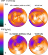
Background: Patient movement has been considered an important source of errors in cardiac PET. This study was aimed at evaluating the effects of such movement on myocardial blood flow (MBF) and perfusable tissue fraction (PTF) measurements in intravenous ¹⁵O-water PET.
Methods: Nineteen ¹⁵O-water scans were performed on ten healthy volunteers and three patients with severe cardiac dysfunction under resting conditions. Motions of subjects during scans were estimated by monitoring locations of markers on their chests using an optical motion-tracking device. Each sinogram of the dynamic emission frames was corrected for subject motion. Variation of regional MBF and PTF with and without the motion corrections was evaluated.
Results: In nine scans, motions during ¹⁵O-water scan (inter-frame (IF) motion) and misalignments relative to the transmission scan (inter-scan (IS) motion) larger than the spatial resolution of the PET scanner (4.0 mm) were both detected by the optical motion-tracking device. After correction for IF motions, MBF values changed from 0.845 ± 0.366 to 0.780 ± 0.360 mL/minute/g (P < .05). In four scans with only IS motion detected, PTF values changed significantly from 0.465 ± 0.118 to 0.504 ± 0.087 g/mL (P< .05), but no significant change was found in MBF values.
Conclusions: This study demonstrates that IF motion during ¹⁵O-water scan at rest can be source of error in MBF measurement. Furthermore, estimated MBF is less sensitive than PTF values to misalignment between transmission and ¹⁵O-water emission scans.
Figures


References
-
- McCord ME, Bacharach SL, Bonow RO, Dilsizian V, Cuocolo A, Freedman N. Misalignment between PET transmission and emission scans: Its effect on myocardial imaging. J Nucl Med. 1992;33:1209–1214. - PubMed
-
- Bacharach SL, Douglas MA, Carson RE, Kalkowski PJ, Freedman NM, Perrone-Filardi P, et al. Three-dimensional registration of cardiac positron emission tomography attenuation scans. J Nucl Med. 1993;34:311–321. - PubMed
-
- Iida H, Kanno I, Takahashi A, Miura S, Murakami M, Takahashi K, et al. Measurement of absolute myocardial blood flow with H215O and dynamic positron-emission tomography. Strategy for quantification in relation to the partial-volume effect. Circulation. 1988;78:104–115. doi: 10.1161/01.CIR.78.1.104. - DOI - PubMed
-
- Iida H, Rhodes CG, de Silva R, Yamamoto Y, Araujo LI, Maseri A, et al. Myocardial tissue fraction-correction for partial volume effects and measure of tissue viability. J Nucl Med. 1991;32:2169–2175. - PubMed
Publication types
MeSH terms
Substances
LinkOut - more resources
Full Text Sources
Medical
Miscellaneous

