. 2012 Mar;61(3):692-701.
doi: 10.2337/db11-1027. Epub 2012 Feb 7.

Age-related impairment in insulin release: the essential role of β(2)-adrenergic receptor

Affiliations

Age-related impairment in insulin release: the essential role of β(2)-adrenergic receptor

Gaetano Santulli et al. Diabetes. 2012 Mar.

Abstract

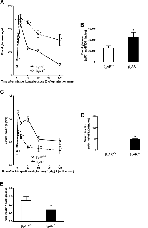
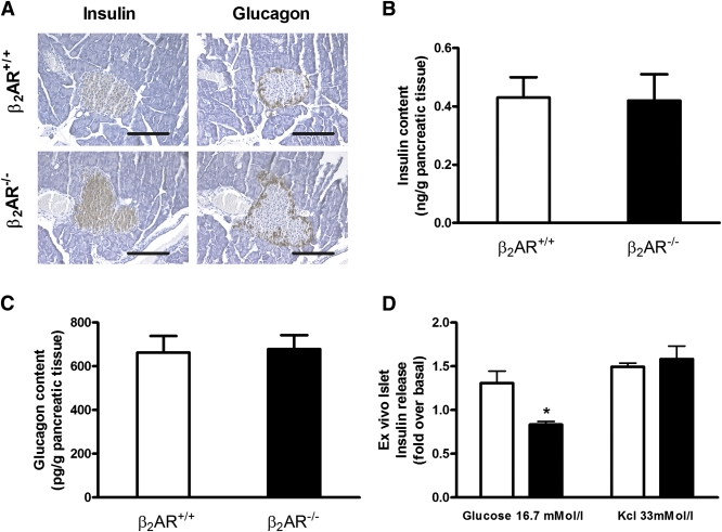
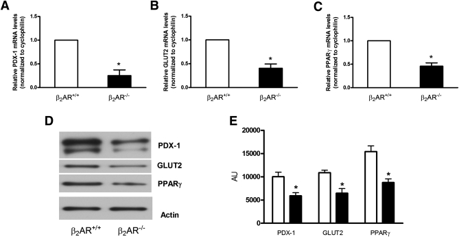
In this study, we investigated the significance of β(2)-adrenergic receptor (β(2)AR) in age-related impaired insulin secretion and glucose homeostasis. We characterized the metabolic phenotype of β(2)AR-null C57Bl/6N mice (β(2)AR(-/-)) by performing in vivo and ex vivo experiments. In vitro assays in cultured INS-1E β-cells were carried out in order to clarify the mechanism by which β(2)AR deficiency affects glucose metabolism. Adult β(2)AR(-/-) mice featured glucose intolerance, and pancreatic islets isolated from these animals displayed impaired glucose-induced insulin release, accompanied by reduced expression of peroxisome proliferator-activated receptor (PPAR)γ, pancreatic duodenal homeobox-1 (PDX-1), and GLUT2. Adenovirus-mediated gene transfer of human β(2)AR rescued these defects. Consistent effects were evoked in vitro both upon β(2)AR knockdown and pharmacologic treatment. Interestingly, with aging, wild-type (β(2)AR(+/+)) littermates developed impaired insulin secretion and glucose tolerance. Moreover, islets from 20-month-old β(2)AR(+/+) mice exhibited reduced density of β(2)AR compared with those from younger animals, paralleled by decreased levels of PPARγ, PDX-1, and GLUT2. Overexpression of β(2)AR in aged mice rescued glucose intolerance and insulin release both in vivo and ex vivo, restoring PPARγ/PDX-1/GLUT2 levels. Our data indicate that reduced β(2)AR expression contributes to the age-related decline of glucose tolerance in mice.


Figures

FIG. 1.
FIG. 1.
Metabolic profile of β2AR−/− mice. Six-month-old β2AR−/− mice and their wild-type littermates (β2AR+/+) were fasted for 16 h and subjected to intraperitoneal glucose loading (2 g/kg body weight). Blood glucose (A and B) and serum insulin (C and D) were monitored for 120 min after glucose administration (n = 14–18 animals per group). β2AR−/− mice displayed glucose intolerance (A) and impaired insulin secretion (C). We calculated the AUC from glucose (B) and insulin excursion (D) curves. Peak insulin–to–peak glucose ratio (E) represents β-cell function, as better described in research design and methods. Bars represent means ± SE. *P < 0.05 vs. β2AR+/+, Bonferroni post hoc test. AUC, area under the curve.
FIG. 2.
FIG. 2.
Comparison of β2AR+/+ and β2AR −/− pancreatic islets. Immunoistochemical analysis (A) of the islets was carried out on paraffin sections using insulin (left panel) or glucagon (right panel) antibodies. Microphotographs are representative of images obtained from pancreas sections of five 6-month-old β2AR+/+ (upper panel) or β2AR −/− (lower panel) mice. Insulin (B) and glucagon (C) content in isolated islets from β2AR+/+ (n = 10) or β2AR −/− (n = 13) mice. Insulin secretion in response to basal (2.8 mmol/L) or high (16.7 mmol/L) glucose concentration and to KCl (33 mmol/L) was measured in isolated islets from β2AR+/+ (□) and β2AR−/− (■) mice (D). Bars represent means ± SE of data from 10 mice per group. *P < 0.05 vs. β2AR+/+, Bonferroni post hoc test. (See also Supplementary Fig. 1.) (A high-quality digital representation of this figure is available in the online issue.)
FIG. 3.
FIG. 3.
Gene expression profile in isolated Langerhans islets from β2AR+/+ and β2AR−/− mice. The abundance of mRNAs for PDX-1 (A), GLUT2 (B), and PPARγ (C) was determined by real-time RT-PCR analysis of total RNA, using cyclophilin as internal standard. The mRNA levels in β2AR−/− mice are relative to those in control animals. Each bar represents means ± SE of four independent experiments in each of which reactions were performed in triplicate using the pooled total RNAs from five mice/genotype. Proteins from a Western blot representative of three independent experiments were quantified by densitometry (D and E). *P < 0.05 vs. β2AR+/+, Bonferroni post hoc test. AU, arbitrary units.
FIG. 4.
FIG. 4.
β2AR levels, glucose-stimulated insulin secretion, and gene expression profile in silenced INS-1E β-cells. Treatment with a specific β2AR-shRNA significantly decreased the density (by 73.7% [A]) and mRNA levels (by 59.1% [B]) of β2AR in INS-1E β-cells. β2AR-shRNA inhibited the insulin secretory response to 16.7 mmol/L glucose, which was rescued by the overexpression of PPARγ (C). KCl-induced insulin release (C) was not significantly different among the studied groups. β2AR-shRNA also determined a significant reduction in mRNA level of PDX-1 (D), GLUT2 (E), and PPARγ (F) that was prevented by the overexpression of PPARγ. Bars represent means ± SE from four to five independent experiments in each of which reactions were performed in triplicate (formula image
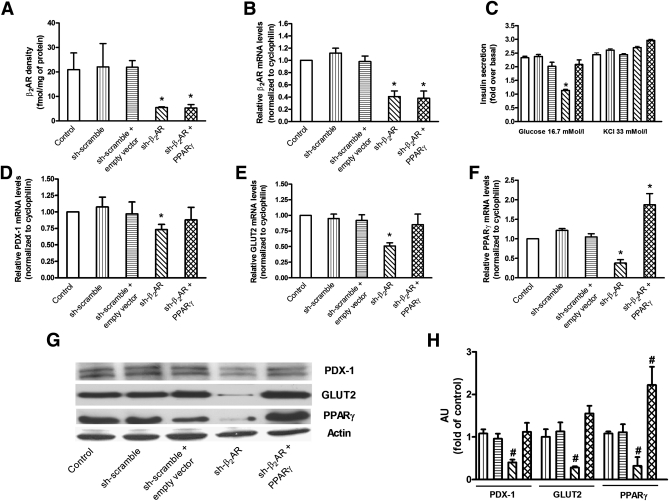
, control, i.e. untreated INS-1E β-cells; formula image, sh-scramble; formula image, sh-scramble+empty vector; formula image, sh-β2AR; formula image, sh-β2AR+PPARγ; *P < 0.05 vs. control, Bonferroni post hoc test; basal is glucose 2.8 mmol/L. Equal amount of proteins from three independent experiments was analyzed by Western blotting and quantified by densitometry (G and H ). *P < 0.05 vs. sh-scramble. AU, arbitrary units. (See also Supplementary Figs. 2–4.)
FIG. 5.
FIG. 5.
β2AR ex vivo infection rescued age-dependent impairment of β-cell function. Density (A) and mRNA levels (B) of β2AR were evaluated on cell membranes of islets isolated from β2AR+/+ mice. Insulin release (C) was determined upon exposure to the indicated concentration of glucose or KCl as described in research design and methods. mRNA levels of PDX-1 (D), GLUT2 (E), and PPARγ (F) were determined by real-time RT-PCR using the pooled total RNAs from five mice/group with cyclophilin as internal standard. Each bar represents means ± SE of five independent experiments in each of which reactions were performed in triplicate. Islets isolated from β2AR+/+ mice were solubilized and aliquots of the lysates were blotted with PDX-1, GLUT2, and PPARγ antibodies. Actin was used as loading control. The autoradiographs shown (G) are representative of three independent experiments, which are quantified in H. formula image
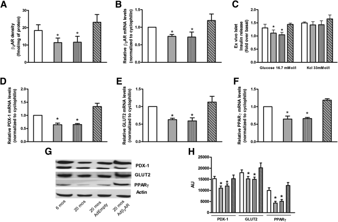
, age 6 months (mos); formula image, 20 months; formula image, 20 months AdEmpty; formula image, 20 months Adβ2AR. *P < 0.05 vs. β2AR+/+ 6 months, Bonferroni post hoc test.
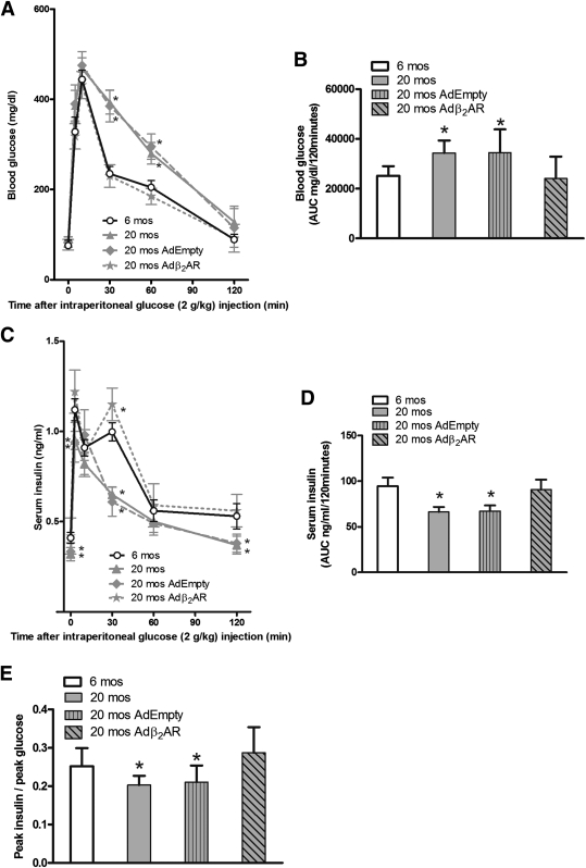
FIG. 6.
FIG. 6.
Adenoviral vector-mediated β2AR gene transfer in the mouse pancreas rescued age-related reduction in glucose tolerance. Blood glucose levels (A) and serum insulin (C) after 120 min of glucose administration (n = 14–18 animals per group). We calculated the AUC from glucose (B) and insulin excursion (D) curves. Twenty-month-old β2AR+/+ mice showed glucose intolerance (A and B), impaired insulin secretion (C and D), and also an impairment in β-cell function, evaluated measuring the peak insulin–to–peak glucose ratio (E). All of these parameters were restored after Adβ2AR in vivo infection. *P < 0.05 vs. β2AR+/+ at 6 months (mos) of age, Bonferroni post hoc test. (See also Supplementary Figs. 5 and 6.)

References

    1. Defronzo RA. Banting Lecture. From the triumvirate to the ominous octet: a new paradigm for the treatment of type 2 diabetes mellitus. Diabetes 2009;58:773–795 - PMC - PubMed
    1. Basu R, Breda E, Oberg AL, et al. Mechanisms of the age-associated deterioration in glucose tolerance: contribution of alterations in insulin secretion, action, and clearance. Diabetes 2003;52:1738–1748 - PubMed
    1. Gumbiner B, Polonsky KS, Beltz WF, Wallace P, Brechtel G, Fink RI. Effects of aging on insulin secretion. Diabetes 1989;38:1549–1556 - PubMed
    1. Ma X, Becker D, Arena VC, Vicini P, Greenbaum C. The effect of age on insulin sensitivity and insulin secretion in first-degree relatives of type 1 diabetic patients: a population analysis. J Clin Endocrinol Metab 2009;94:2446–2451 - PMC - PubMed
    1. Reaven E, Wright D, Mondon CE, Solomon R, Ho H, Reaven GM. Effect of age and diet on insulin secretion and insulin action in the rat. Diabetes 1983;32:175–180 - PubMed