. 2012 Feb 21;109(8):3012-7.
doi: 10.1073/pnas.1117359109. Epub 2012 Feb 6.

Plasmacytoid dendritic cells control T-cell response to chronic viral infection

Affiliations

Plasmacytoid dendritic cells control T-cell response to chronic viral infection

Luisa Cervantes-Barragan et al. Proc Natl Acad Sci U S A. .

Abstract

Infections with persistent viruses are a frequent cause of immunosuppression, autoimmune sequelae, and/or neoplastic disease. Plasmacytoid dendritic cells (pDCs) are innate immune cells that produce type I interferon (IFN-I) and other cytokines in response to virus-derived nucleic acids. Persistent viruses often cause depletion or functional impairment of pDCs, but the role of pDCs in the control of these viruses remains unclear. We used conditional targeting of pDC-specific transcription factor E2-2 to generate mice that constitutively lack pDCs in peripheral lymphoid organs and tissues. The profound impact of pDC deficiency on innate antiviral responses was revealed by the failure to control acute infection with the cytopathic mouse hepatitis virus. Furthermore, pDC-deficient animals failed to clear lymphocytic choriomeningitis virus (LCMV) from hematopoietic organs during persistent LCMV infection. This failure was associated with reduced numbers and functionality of LCMV-specific CD4(+) helper T cells and impaired antiviral CD8(+) T-cell responses. Adoptive transfer of LCMV-specific T cells revealed that both CD4(+) and CD8(+) T cells required IFN-I for expansion, but only CD4(+) T cells required the presence of pDCs. In contrast, mice with pDC-specific loss of MHC class II expression supported normal CD4(+) T-cell response to LCMV. These data suggest that pDCs facilitate CD4(+) helper T-cell responses to persistent viruses independently of direct antigen presentation. Thus pDCs provide an essential link between innate and adaptive immunity to chronic viral infection, likely through the secretion of IFN-I and other cytokines.


Conflict of interest statement

The authors declare no conflict of interest.

Figures

Fig. 1.
Fig. 1.
A mouse model of constitutive pDC ablation. CKO animals with DC-specific E2-2 deletion (Tcf4flox/− Itgax-Cre+) were analyzed in parallel to Tcf4flox/+ Itgax-Cre littermate controls (Ctrl). Statistically significant differences are indicated as follows: ***P < 0.001; **P < 0.01; *P < 0.05. (A) The pDC population in CKO mice. Shown are representative staining profiles of the bone marrow (BM) or splenic cells, with the frequencies of Bst2+ CD11clow pDC population indicated (mean ± SD of three or four animals for the bone marrow and nine animals for the spleen). (B) The cDC population in CKO mice. Shown are staining profiles of total splenocytes with the CD11chi MHC class II+ (MHCII) cDC subset highlighted and the two cDC subsets within the gated cDC population. Subset frequencies represent mean ± SD of seven animals. The increase of the cDC fraction in CKO spleens is significant (P = 0.006). The B220+ CD8+ cDC-like cells arising from E2-2–deficient peripheral pDCs (28) could not be detected reliably in CKO mice. (C) Absolute numbers of pDCs and cDCs in lymphoid organs. Data shown are mean ± SD of four animals. (D) IFN-α production by total bone marrow cells or splenocytes cultured for 48 h with CpG DNA. Shown are IFN-α concentrations in the supernatant (mean ± SD of three to five animals). (E) IFN-α production after in vivo challenge with CpG DNA. Shown are serum IFN-α concentrations 6 h after CpG injection (mean ± SD of three animals).
Fig. 2.
Fig. 2.
The loss of pDCs abolishes innate control of acute MHV infection. Groups of five control and five CKO mice were infected with 50 pfu MHV A59 and analyzed 48 h later. Statistical significance is indicated as in Fig. 1. (A) Serum IFN-α concentrations determined by ELISA (mean ± SD). (B) Virus titers in the indicated organs (mean ± SD). (C) Serum levels of ALT indicative of liver damage (mean ± SD). (D) Representative liver sections stained with H&E. Arrows indicate inflammatory foci in livers of CKO mice. (Scale bar, 100 μm.)
Fig. 3.
Fig. 3.
Persistence of LCMV Docile in the blood of pDC-deficient mice. CKO mice or littermate controls were infected with 105 pfu of LCMV Docile, and viral titers in blood were determined at the indicated time points postinfection. Data shown are mean ± SD of six mice per group.
Fig. 4.
Fig. 4.
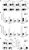
Impaired T-cell response to chronic LCMV infection in the absence of pDCs. CKO mice or littermate controls were infected with 105 pfu of LCMV Docile, and LCMV-specific T-cell responses were analyzed on days 7 or 30. Total splenocytes were stained with LCMV peptide-MHC class I tetramers (tet) or were stimulated with LCMV peptides and stained for intracellular IFN-γ. Statistical significance is indicated as in Fig. 1. (A) Representative staining profiles of splenocytes highlighting gp33-specific CD8+ T cells and gp33-responsive IFN-γ–producing CD8+ T cells. (B and C) Frequencies of tetramer-positive CD8+ T cells (B) and of IFN-γ–producing CD8+ T cells (C) specific for the three indicated LCMV peptides. Data shown are mean ± SD of 8–11 mice pooled from three independent experiments. (D) Representative staining profiles of splenocytes highlighting gp61-responsive IFN-γ–producing CD4+ T cells. Values in the upper right quadrant indicate the percentage of IFN-γ–producing cells in the CD4+ T cell compartment. (E) The frequency of gp61-responsive IFN-γ–producing CD4+ T cells. Data shown are mean ± SD of seven mice from two independent experiments. p.i., postinfection.
Fig. 5.
Fig. 5.
The absence of pDCs impairs CD4+ T-cell response to chronic LCMV infection. The indicated mice were infected with 105 pfu of LCMV Docile, received adoptive transfer of LCMV peptide gp61-specific SMARTA1 (SM1) TCR transgenic CD4+ T cells on day 1, and were analyzed on day 8. Statistical significance is indicated as in Fig. 1. (A) Expansion of the transferred SM1 TCR transgenic CD4+ T cells in Tlr7-deficient mice. Shown is the fraction of Thy1.1+ transferred T cells among the splenic CD4+ T-cell population in Tlr7−/− or control C57BL/6 (B6) mice (mean ± SD; n = 5). (B) Expansion of SM1 T cells in pDC-deficient CKO or control (Ctrl) mice (mean ± SD; n = 8). (C) Surface levels of PD-1 and BTLA on the transferred T cells, with the mean fluorescence intensities indicated (mean ± SD; n = 8). (D) Expansion of IFNAR-deficient TCR transgenic CD4+ T cells. A 1:1 mixture of wild-type (Thy1.1+ Ly5.1) and IFNAR-deficient (Thy1.1 Ly5.1+) SM1 T cells was transferred into LCMV-infected control and CKO mice. Shown are fractions of transferred T cells among the splenic CD4+ T-cell population on day 8 (mean ± SD; n = 8). (E) Cytokine production by wild-type SM1 T cells transferred into LCMV-infected control or CKO recipients. Splenocytes were stimulated with gp61 peptide 8 d after the transfer. Shown is the frequency of cells producing indicated cytokines among the transferred Thy1.1+ SM1 T cells (mean ± SD; n = 6). (F) Expansion of SM1 cells in mice with pDC-specific deletion of MHC class II. The experiment was done as in D, with SM1 T cells transferred into LCMV-infected control (μMT:WT) or pDC-specific MHC class II knockout (μMT×PIII+IV-KO:WT) mice. Shown are fractions of transferred T cells among the splenic CD4+ T-cell population on day 8 (mean ± SD; n = 4). (G) Production of IFN-γ by wild-type SM1 T cells transferred into LCMV-infected control (μMT:WT) or pDC-specific MHC class II knockout (μMT×PIII+IV-KO:WT) mice. The results are shown as in E (mean ± SD; n = 4).
Fig. 6.
Fig. 6.
The absence of pDCs does not directly affect CD8+ T-cell response to chronic LCMV infection. CKO or control mice were infected with 105 pfu of LCMV Docile, received adoptive transfer of LCMV peptide gp33-specific P14 TCR transgenic CD8+ T cells on day 1, and were analyzed on day 8. Data represent eight animals per group pooled from two independent experiments. Statistical significance is indicated as in Fig. 1. (A) Expansion of the transferred P14 TCR transgenic CD8+ T cells. Shown is the fraction of Ly5.1+ transferred T cells among the splenic CD8+ T-cell population in control or CKO mice (mean ± SD). (B) Surface levels of PD-1 and BTLA on the transferred T cells, with the mean fluorescence intensities indicated. (C) Expansion of IFNAR-deficient TCR transgenic CD8+ T cells. A 1:1 mixture of wild-type (Thy1.1 Ly5.1+) and IFNAR-deficient (Thy1.1+ Ly5.1) TCR transgenic CD8+ T cells was transferred into LCMV-infected control and CKO mice. Shown are fractions of transferred T cells among the splenic CD8+ T-cell population on day 8 (mean ± SD).

References

    1. Klenerman P, Hill A. T cells and viral persistence: Lessons from diverse infections. Nat Immunol. 2005;6:873–879. - PubMed
    1. Buchmeier MJ, Welsh RM, Dutko FJ, Oldstone MB. The virology and immunobiology of lymphocytic choriomeningitis virus infection. Adv Immunol. 1980;30:275–331. - PubMed
    1. Moskophidis D, Lechner F, Hengartner H, Zinkernagel RM. MHC class I and non-MHC-linked capacity for generating an anti-viral CTL response determines susceptibility to CTL exhaustion and establishment of virus persistence in mice. J Immunol. 1994;152:4976–4983. - PubMed
    1. Oldstone MB. Anatomy of viral persistence. PLoS Pathog. 2009;5:e1000523. - PMC - PubMed
    1. Bergthaler A, et al. Viral replicative capacity is the primary determinant of lymphocytic choriomeningitis virus persistence and immunosuppression. Proc Natl Acad Sci USA. 2010;107:21641–21646. - PMC - PubMed

Publication types

MeSH terms

Substances