. 2012 Feb 21;109(8):3137-42.
doi: 10.1073/pnas.1200718109. Epub 2012 Feb 6.

Polycation-siRNA nanoparticles can disassemble at the kidney glomerular basement membrane

Affiliations

Polycation-siRNA nanoparticles can disassemble at the kidney glomerular basement membrane

Jonathan E Zuckerman et al. Proc Natl Acad Sci U S A. .

Abstract

Despite being engineered to avoid renal clearance, many cationic polymer (polycation)-based siRNA nanoparticles that are used for systemic delivery are rapidly eliminated from the circulation. Here, we show that a component of the renal filtration barrier--the glomerular basement membrane (GBM)--can disassemble cationic cyclodextrin-containing polymer (CDP)-based siRNA nanoparticles and, thereby, facilitate their rapid elimination from circulation. Using confocal and electron microscopies, positron emission tomography, and compartment modeling, we demonstrate that siRNA nanoparticles, but not free siRNA, accumulate and disassemble in the GBM. We also confirm that the siRNA nanoparticles do not disassemble in blood plasma in vitro and in vivo. This clearance mechanism may affect any nanoparticles that assemble primarily by electrostatic interactions between cationic delivery components and anionic nucleic acids (or other therapeutic entities).


Conflict of interest statement

The authors declare no conflict of interest.

Figures

Fig. 1.
Fig. 1.
Characterization of siRNA nanoparticles. (A) siRNA nanoparticles assemble because of electrostatic interactions between the cationic cyclodextrin containing polymer and the anionic siRNA. PEG provides steric stabilization and is bound to the particles via inclusion complex formation between its terminal adamantane (AD) modification and the cyclodextrin cup of the CDP. (B) Cryo-TEM images of siRNA nanoparticles revealed sub–100-nm spherical objects. (Scale bars: 100 nm.) (C) Nanoparticle tracking analysis of siRNA nanoparticle sizes and zeta potentials (error bars represent SD of three measurements, n = 3).
Fig. 2.
Fig. 2.
Nanoparticle components remained associated and assembled in vivo but were disassembled by heparan sulfate. (AC) Gel mobility shift assays demonstrated siRNA/CDP association. Free siRNA will migrate down the gel toward the anode, whereas siRNA/CDP nanoparticles remain in the wells or migrated toward the cathode. (A) In vitro siRNA formulations were formulated as indicated (formulated nanoparticles, siRNA+AD-PEG/CDP in H2O) and incubated at 37 °C for 15 min. Plasma samples: Formulated nanoparticles, plasma from animals 3 min after injection of formulated siRNA+AD-PEG/CDP nanoparticles. Sequential injection of components, plasma from animals where free siRNA was injected and then 1 min later CDP/AD-PEG were injected; plasma was collected at 3 min after the first injection. siRNA or CDP only, plasma collected from animals 3 min after receiving injection of only siRNA or AD-PEG/CDP. Disassembled nanoparticles, plasma from animals where formulated siRNA nanoparticles were injected and then 1 min later an excess of ≈6 kb of plasmid DNA was injected, plasma was collected at 3 min after the first injection. All duplicate lanes are from independent animals. (B) Plasma samples from animals receiving formulated siRNA nanoparticles taken at the indicated time point after injection. (C) Gel mobility shift assays of siRNA nanoparticles in increasing amounts of heparan sulfate in 50% (vol/vol) mouse plasma. All plasma containing samples have a band of background staining that migrates at ≈5 kb as indicated in the figure.
Fig. 3.
Fig. 3.
Real-time PET imaging and compartment model of GBM-induced disassembly of siRNA nanoparticles. (A) Images of PET signal from kidneys and bladder of mice receiving free and nanoparticle-formulated 64Cu-DOTA–labeled siRNA (data adapted from ref. 10). (B) Quantification of kidney, blood, and bladder 64Cu-DOTA labeled siRNA intensities from PET studies [Error bars = SD, free siRNA n = 4, siRNA nanoparticles (NPs), n = 5]. (C) Computed results from compartment model of PET data for free siRNA (red) and siRNA nanoparticles (black).
Fig. 4.
Fig. 4.
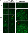
siRNA nanoparticles, but not free siRNA, transiently accumulate in glomeruli after i.v. administration. Time course of confocal microcopy images of kidneys extracted from mice receiving free Cy3-labeled siRNA (A), Cy3-labled siRNA nanoparticles (B), or no treatment (C). Higher magnification images of glomeruli from 6 min (D) and 10 min (E) time points. White arrows indicate glomeruli positions, blue arrows indicate areas of tubular Cy3-signal accumulation, and yellow arrows indicate cy3 fluorescence in peri-tubule vasculature lining.
Fig. 5.
Fig. 5.
Nanoparticles accumulate and disassemble at the kidney glomerular basement membrane. (A) Image of GBM from an animal receiving only free siRNA. (B) Low magnification EM image of glomerular capillaries from a mouse 10 min after i.v. administration of siRNA nanoparticles. (C and D) Higher magnification images of the GBM regions of these glomerular capillaries. BM, Basement membrane; E, Endothelial cell; FB, Filtration barrier, M, Mesangium, (I/D)-NP, (intact/disassembling) nanoparticle, P, podoyctes; PC, peritubule capillary; PF, podocyte foot process; R, Erythrocyte; U, Urinary space.
Fig. 6.
Fig. 6.
Schematic of siRNA nanoparticle deposition and disassembly in the GBM with key modeling expressions highlighted. Nanoparticles cross through fenestrations in the glomerular endothelial cell lining and enter the GBM. Within the GBM, the nanoparticles are disassembled by the abundant heparan sulfate molecules. Once disassembled, the nanoparticle components can cross the remainder of the GBM and the podocyte filtration slits and enter the urinary space.

References

    1. Castanotto D, Rossi JJ. The promises and pitfalls of RNA-interference-based therapeutics. Nature. 2009;457:426–433. - PMC - PubMed
    1. de Fougerolles A, Vornlocher HP, Maraganore J, Lieberman J. Interfering with disease: A progress report on siRNA-based therapeutics. Nat Rev Drug Discov. 2007;6:443–453. - PMC - PubMed
    1. Davis ME, Chen ZG, Shin DM. Nanoparticle therapeutics: An emerging treatment modality for cancer. Nat Rev Drug Discov. 2008;7:771–782. - PubMed
    1. Davis ME, et al. Evidence of RNAi in humans from systemically administered siRNA via targeted nanoparticles. Nature. 2010;464:1067–1070. - PMC - PubMed
    1. de Wolf HK, et al. Effect of cationic carriers on the pharmacokinetics and tumor localization of nucleic acids after intravenous administration. Int J Pharm. 2007;331:167–175. - PubMed

Publication types

MeSH terms

LinkOut - more resources