

The impact of cyclin D1 mRNA isoforms, morphology and p53 in mantle cell lymphoma: p53 alterations and blastoid morphology are strong predictors of a high proliferation index
- PMID: 22315488
- PMCID: PMC3436245
- DOI: 10.3324/haematol.2011.055715
The impact of cyclin D1 mRNA isoforms, morphology and p53 in mantle cell lymphoma: p53 alterations and blastoid morphology are strong predictors of a high proliferation index
Abstract
Background: Mantle cell lymphoma is a clinically heterogeneous disease characterized by overexpression of cyclin D1 protein. Blastoid morphology, high proliferation, and secondary genetic aberrations are markers of aggressive behavior. Expression profiling of mantle cell lymphoma revealed that predominance of the 3'UTR-deficient, short cyclin D1 mRNA isoform was associated with high cyclin D1 levels, a high "proliferation signature" and poor prognosis.
Design and methods: Sixty-two cases of mantle cell lymphoma were analyzed for cyclin D1 mRNA isoforms and total cyclin D1 levels by real-time reverse transcriptase polymerase chain reaction, and TP53 alterations were assessed by immunohistochemistry and molecular analysis. Results were correlated with proliferation index and clinical outcome.
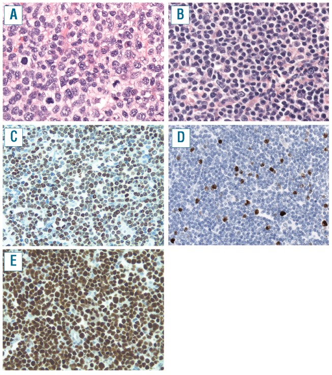
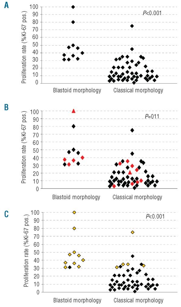
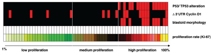
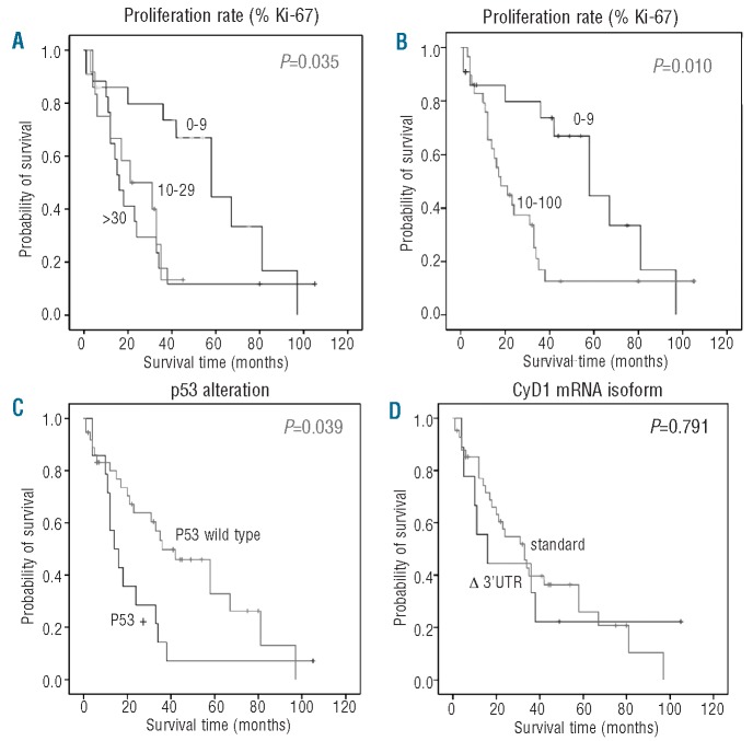
Results: Predominance of the short cyclin D1 mRNA was found in 14 (23%) samples, including four with complete loss of the standard transcript. TP53 alterations were found in 15 (24%) cases. Predominance of 3'UTR-deficient mRNA was significantly associated with high cyclin D1 mRNA levels (P=0.009) and more commonly found in blastoid mantle cell lymphoma (5/11, P=0.060) and cases with a proliferation index of >20% (P=0.026). Both blastoid morphology (11/11, P<0.001) and TP53 alterations (15/15, P<0.001) were significantly correlated with a high proliferation index. A proliferation index of 10% was determined to be a significant threshold for survival in multivariate analysis (P=0.01).
Conclusions: TP53 alterations are strongly associated with a high proliferation index and aggressive behavior in mantle cell lymphoma. Predominance of the 3'UTR-deficient transcript correlates with higher cyclin D1 levels and may be a secondary contributing factor to high proliferation, but failed to reach prognostic significance in this study.
Figures
References
-
- Bosch F, Jares P, Campo E, Lopez-Guillermo A, Piris MA, Villamor N, et al. PRAD-1/cyclin D1 gene overexpression in chronic lymphoproliferative disorders: a highly specific marker of mantle cell lymphoma. Blood. 1994;84(8):2726–32. - PubMed
-
- Campo E, Raffeld M, Jaffe ES. Mantle-cell lymphoma. Semin Hematol. 1999;36(2):115–27. - PubMed
-
- Bergsagel PL, Kuehl WM. Critical roles for immunoglobulin translocations and cyclin D dysregulation in multiple myeloma. Immunol Rev. 2003;194:96–104. - PubMed
-
- de Boer CJ, Kluin-Nelemans JC, Dreef E, Kester MG, Kluin PM, Schuuring E, et al. Involvement of the CCND1 gene in hairy cell leukemia. Ann Oncol. 1996;7(3):251–6. - PubMed
Publication types
MeSH terms
Substances
LinkOut - more resources
Full Text Sources
Research Materials
Miscellaneous
