

Amniotic mesenchymal stem cells: a new source for hepatocyte-like cells and induction of CFTR expression by coculture with cystic fibrosis airway epithelial cells
- PMID: 22315512
- PMCID: PMC3270433
- DOI: 10.1155/2012/575471
Amniotic mesenchymal stem cells: a new source for hepatocyte-like cells and induction of CFTR expression by coculture with cystic fibrosis airway epithelial cells
Abstract
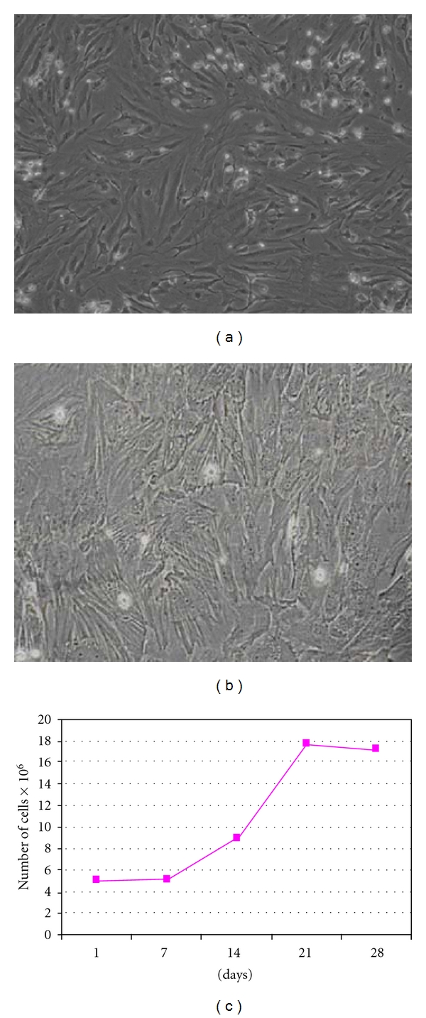
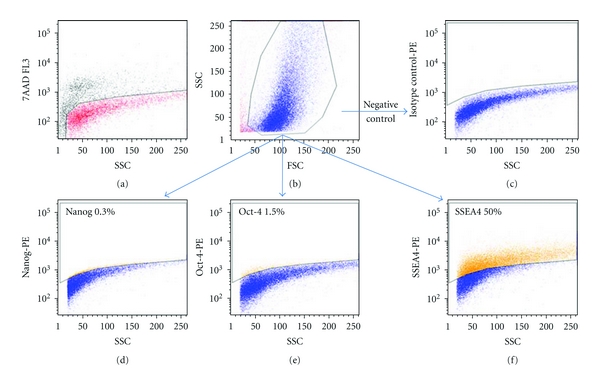
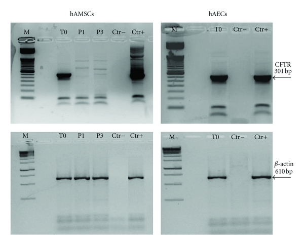
Cystic fibrosis (CF) is a monogenic disease caused by mutations in the CF transmembrane conductance regulator (CFTR) gene, with lung and liver manifestations. Because of pitfalls of gene therapy, novel approaches for reconstitution of the airway epithelium and CFTR expression should be explored. In the present study, human amniotic mesenchymal stem cells (hAMSCs) were isolated from term placentas and characterized for expression of phenotypic and pluripotency markers, and for differentiation potential towards mesoderm (osteogenic and adipogenic) lineages. Moreover, hAMSCs were induced to differentiate into hepatocyte-like cells, as demonstrated by mixed function oxidase activity and expression of albumin, alpha1-antitrypsin, and CK19. We also investigated the CFTR expression in hAMSCs upon isolation and in coculture with CF airway epithelial cells. Freshly isolated hAMSCs displayed low levels of CFTR mRNA, which even decreased with culture passages. Following staining with the vital dye CM-DiI, hAMSCs were mixed with CFBE41o- respiratory epithelial cells and seeded onto permeable filters. Flow cytometry demonstrated that 33-50% of hAMSCs acquired a detectable CFTR expression on the apical membrane, a result confirmed by confocal microscopy. Our data show that amniotic MSCs have the potential to differentiate into epithelial cells of organs relevant in CF pathogenesis and may contribute to partial correction of the CF phenotype.
Figures


Similar articles
-
Correction of defective CFTR/ENaC function and tightness of cystic fibrosis airway epithelium by amniotic mesenchymal stromal (stem) cells.J Cell Mol Med. 2014 Aug;18(8):1631-43. doi: 10.1111/jcmm.12303. Epub 2014 Jun 3. J Cell Mol Med. 2014. PMID: 24894806 Free PMC article.
-
CFTR delivery to 25% of surface epithelial cells restores normal rates of mucus transport to human cystic fibrosis airway epithelium.PLoS Biol. 2009 Jul;7(7):e1000155. doi: 10.1371/journal.pbio.1000155. Epub 2009 Jul 21. PLoS Biol. 2009. PMID: 19621064 Free PMC article.
-
Transient receptor potential canonical channel 6 links Ca2+ mishandling to cystic fibrosis transmembrane conductance regulator channel dysfunction in cystic fibrosis.Am J Respir Cell Mol Biol. 2011 Jan;44(1):83-90. doi: 10.1165/rcmb.2009-0347OC. Epub 2010 Mar 4. Am J Respir Cell Mol Biol. 2011. PMID: 20203293
-
Correction of Airway Stem Cells: Genome Editing Approaches for the Treatment of Cystic Fibrosis.Hum Gene Ther. 2020 Sep;31(17-18):956-972. doi: 10.1089/hum.2020.160. Epub 2020 Sep 8. Hum Gene Ther. 2020. PMID: 32741223 Free PMC article. Review.
-
Emerging relationship between CFTR, actin and tight junction organization in cystic fibrosis airway epithelium.Histol Histopathol. 2017 May;32(5):445-459. doi: 10.14670/HH-11-842. Epub 2016 Nov 11. Histol Histopathol. 2017. PMID: 27834058 Review.
Cited by
-
Cell-Based Therapeutic Approaches for Cystic Fibrosis.Int J Mol Sci. 2020 Jul 23;21(15):5219. doi: 10.3390/ijms21155219. Int J Mol Sci. 2020. PMID: 32718005 Free PMC article. Review.
-
The Oncogenic Theory of Preeclampsia: Is Amniotic Mesenchymal Stem Cells-Derived PLAC1 Involved?Int J Mol Sci. 2023 Feb 10;24(4):3612. doi: 10.3390/ijms24043612. Int J Mol Sci. 2023. PMID: 36835024 Free PMC article.
-
Mesenchymal stem or stromal cells from amnion and umbilical cord tissue and their potential for clinical applications.Cells. 2012 Nov 12;1(4):1061-88. doi: 10.3390/cells1041061. Cells. 2012. PMID: 24710543 Free PMC article.
-
Stem cells, cell therapies, and bioengineering in lung biology and diseases. Comprehensive review of the recent literature 2010-2012.Ann Am Thorac Soc. 2013 Oct;10(5):S45-97. doi: 10.1513/AnnalsATS.201304-090AW. Ann Am Thorac Soc. 2013. PMID: 23869446 Free PMC article. Review.
-
Functional Rescue of F508del-CFTR Using Small Molecule Correctors.Front Pharmacol. 2012 Sep 26;3:160. doi: 10.3389/fphar.2012.00160. eCollection 2012. Front Pharmacol. 2012. PMID: 23055971 Free PMC article.
References
-
- Miki T, Strom SC. Amnion-derived pluripotent/multipotent stem cells. Stem Cell Reviews. 2006;2(2):133–142. - PubMed
-
- Parolini O, Alviano F, Bagnara GP, et al. Concise review: isolation and characterization of cells from human term placenta: outcome of the first international workshop on placenta derived stem cells. Stem Cells. 2008;26(2):300–311. - PubMed
-
- Sheppard DN, Welsh MJ. Structure and function of the CFTR chloride channel. Physiological Reviews. 1999;79(1, supplement):S23–S45. - PubMed
-
- Griesenbach U, Alton EWFW. Cystic fibrosis gene therapy: successes, failures and hopes for the future. Expert Review of Respiratory Medicine. 2009;3(4):363–371. - PubMed
