

Isolation of cellulose-degrading bacteria and determination of their cellulolytic potential
- PMID: 22315612
- PMCID: PMC3270400
- DOI: 10.1155/2012/578925
Isolation of cellulose-degrading bacteria and determination of their cellulolytic potential
Abstract
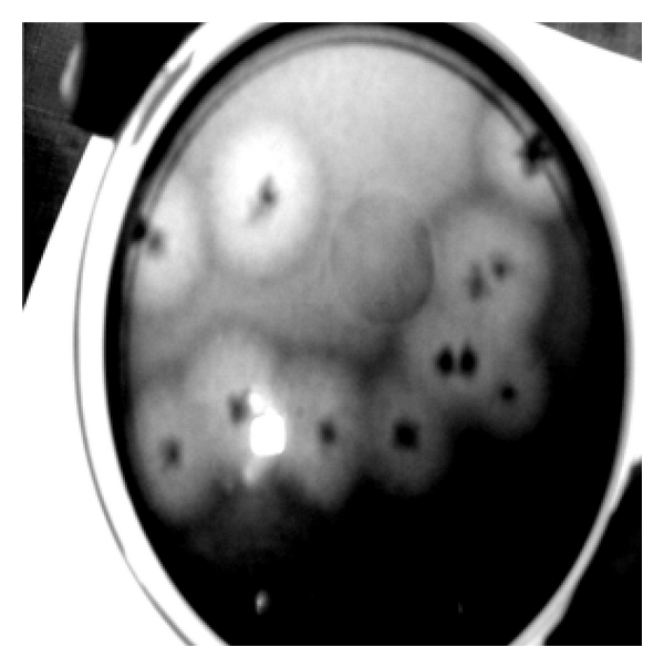
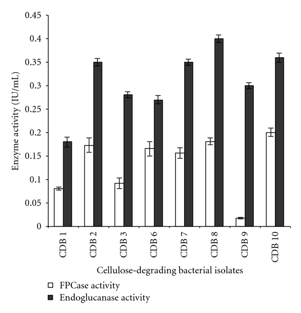
Eight isolates of cellulose-degrading bacteria (CDB) were isolated from four different invertebrates (termite, snail, caterpillar, and bookworm) by enriching the basal culture medium with filter paper as substrate for cellulose degradation. To indicate the cellulase activity of the organisms, diameter of clear zone around the colony and hydrolytic value on cellulose Congo Red agar media were measured. CDB 8 and CDB 10 exhibited the maximum zone of clearance around the colony with diameter of 45 and 50 mm and with the hydrolytic value of 9 and 9.8, respectively. The enzyme assays for two enzymes, filter paper cellulase (FPC), and cellulase (endoglucanase), were examined by methods recommended by the International Union of Pure and Applied Chemistry (IUPAC). The extracellular cellulase activities ranged from 0.012 to 0.196 IU/mL for FPC and 0.162 to 0.400 IU/mL for endoglucanase assay. All the cultures were also further tested for their capacity to degrade filter paper by gravimetric method. The maximum filter paper degradation percentage was estimated to be 65.7 for CDB 8. Selected bacterial isolates CDB 2, 7, 8, and 10 were co-cultured with Saccharomyces cerevisiae for simultaneous saccharification and fermentation. Ethanol production was positively tested after five days of incubation with acidified potassium dichromate.
Figures


References
-
- Shewale JG. Glucosidase: its role in cellulase synthesis and hydrolysis of cellulose. International Journal of Biochemistry. 1982;14(6):435–443. - PubMed
-
- Woodward J, Wiseman A. Fungal and other β-d-glucosidases: their properties and applications. Enzyme and Microbial Technology. 1983;4(2):73–79.
-
- Ryu DDY, Mandels M. Cellulases: biosynthesis and applications. Enzyme and Microbial Technology. 1980;2(2):91–102.
-
- Samdhu DK, Bawa S. Improvement of cellulase activity in Trichoderma . Applied Biochemistry and Biotechnology. 1992;34-35(1):175–192.
LinkOut - more resources
Full Text Sources
Molecular Biology Databases
Research Materials
