. 2011:1:45.
doi: 10.4103/2156-7514.84323. Epub 2011 Aug 27.

Computed tomography findings in xanthogranulomatous pyelonephritis

Affiliations

Computed tomography findings in xanthogranulomatous pyelonephritis

Arumugam Rajesh et al. J Clin Imaging Sci. 2011.

Abstract

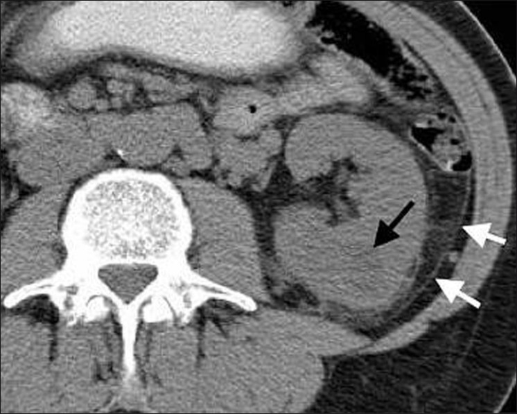
Background: Xanthogranulomatous pyelonephritis (XGN) is an uncommon condition characterized by chronic suppurative renal inflammation that leads to progressive parenchymal destruction.

Purpose: To review the computed tomography (CT) findings of patients diagnosed with XGN.

Materials and methods: A retrospective review of CT findings in patients with histologically proven XGN was carried out.

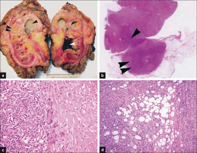
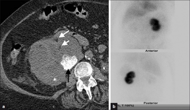
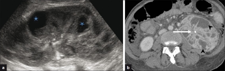
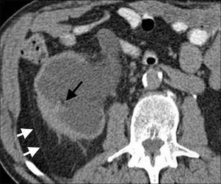
Results: Thirteen CT examinations of 11 patients were analyzed. Renal enlargement was demonstrable on the affected side in all patients. Nine patients (82%) had multiple dilated calyces and abnormal parenchyma. Six patients (55%) had a renal pelvis or upper ureteric calculus causing obstruction. Three patients (27%) had focal fat deposits identifiable within the inflamed renal parenchyma. Two patients had renal abscesses. Ten patients (91%) had extrarenal extension of the inflammatory changes. Three patients (27%) demonstrated extensive retroperitoneal inflammation.

Conclusion: Unilateral renal enlargement and inflammation were the most consistent findings of XGN on CT. Perinephric inflammation and collections or abscess should also alert the radiologist to the possibility of this diagnosis.

Keywords: Xanthogranulomatous pyelonephritis; computed tomography; perinephric inflammation.


Conflict of interest statement

Conflict of Interest: None declared.

Figures

Figure 1
Figure 1
(a) Gross nephrectomy specimen showing characteristic lobulated xanthomatous lesions confined to the renal parenchyma (double arrowhead) caused by an obstructing “staghorn” calculus (white arrowhead) within the renal pelvis which has caused secondary hydronephrosis The dilated upper pole calyces contain purulent green colored fluid (single black arrowhead). (b) H and E section showing focal XGN. Note the wedge-shaped area of cortical inflammation (arrowhead) in continuity with a larger nodular area extending into perinephric fat (double arrowhead). (c) Renal parenchymal granulomatous inflammation with multinucleate giant cells and focal necrosis (far right). The inflammatory infiltrate is rich in histiocytes, some of which have a foamy appearance reflecting high intracellular fat content. (d) The inflammatory reaction extends into perinephric fat (represented by the clear spaces) and with time will undergo organization and fibrosis.
Figure 2
Figure 2
An 85-year-old female presented with weight loss and hypercalcemia. Contrast-enhanced CT shows multiple low attenuation areas (arrow) in right kidney surrounded by intensely enhancing walls. Low attenuation areas represent dilated calyces and pus-filled spaces replacing renal parenchyma.
Figure 3
Figure 3
(a) XGN in an 82-year-old female with known renal stones who presented with obstructive uropathy and hyperkalemia. Noncontrast CT image shows right renal enlargement and dilated calyces with an obstructing renal calculus (black arrow). Also seen are focal xanthomas (white arrows). (b) DMSA image shows no uptake in right kidney and 100% function in left kidney. Histology post nephrectomy confirmed XGN.
Figure 4
Figure 4
(a) XGN in a 40-year-old female with chronic pancreatitis, fever, and weight loss. Ultrasound image showed an enlarged kidney with focal cystic areas (stars), reported as likely renal abscesses. (b) Selected axial CT image shows multiple enhancing dilated calyces (black arrowheads) and an obstructing renal calculus (white arrow).
Figure 5
Figure 5
XGN in an 82-year-old male with perinephric inflammatory changes. There is perinephric stranding and thickening of the posterior pararenal fascia (white arrows). Also seen are focal xanthomas (black arrow).
Figure 6
Figure 6
Focal XGN in a 54-year-old with unexplained weight loss. Low attenuation lesion (23 HU) in lower pole of left kidney (black arrow) Note associated inflammation in the posterior perinephric space and thickened pararenal fascia (white arrows).

References

    1. Kumar V, Abbas A, Fausto N, editors. Robbins and Cotran Pathologic Basis of Disease. 7th ed. Philadelphia, Pa: Saunders Elsevier; 2005.
    1. Hayes WS, Hartman DS, Sesterbenn IA. Xanthogranulomatous pyelonephritis. Radiographics. 1991;11:485–98. - PubMed
    1. Clapton WK, Boucaut HA, Dewan PA, Bourne AJ, Byard RW. Clinicopathological features of xanthogranulomatous pyelonephritis in infancy. Pathology. 1993;25:110–3. - PubMed
    1. Fan CM, Whitman GJ, Chew FS. Xanthogranulomatous pyelonephritis. AJR Am J Roentgenol. 1995;165:1008. - PubMed
    1. Dwivedi US, Goyal NK, Saxena V, Acharya RL, Trivedi S, Singh PB, et al. Xanthogranulomatous pyelonephritis: Our experience with review of published reports. ANZ J Surg. 2006;76:1007–9. - PubMed