Review
. 2012 Jan;2(1):a006429.
doi: 10.1101/cshperspect.a006429.

Endothelial cell heterogeneity

Affiliations
Review

Endothelial cell heterogeneity

William C Aird. Cold Spring Harb Perspect Med. 2012 Jan.

Abstract

The endothelial lining of blood vessels shows remarkable heterogeneity in structure and function, in time and space, and in health and disease. An understanding of the molecular basis for phenotypic heterogeneity may provide important insights into vascular bed-specific therapies. First, we review the scope of endothelial heterogeneity and discuss its proximate and evolutionary mechanisms. Second, we apply these principles, together with their therapeutic implications, to a representative vascular bed in disease, namely, tumor endothelium.


Figures

Figure 1.
Figure 1.
Mechanisms of endothelial cell heterogeneity. (A) Hemangioblasts give rise to endothelial progenitor cells (angioblasts), which in turn differentiate into endothelial cells of arteries, veins, and capillaries. Cell phenotypes are represented by color shades. Shown is the hypothetical relative role of microenvironment and epigenetics in mediating cell type–specific phenotypes. (B) The role of the microenvironment in mediating nonheritable changes in endothelial cell phenotype is represented by receptor-mediated posttranslational modification of protein (e.g., phosphorylation of a signal intermediate) and transcription factor–dependent induction of gene expression. Removal of the extracellular signal will result in eventual loss of translational/transcriptional effects, and residual effects will be “diluted out” with cell division. (C) The role of epigenetics in mediating heritable changes in endothelial cell phenotype is represented by DNA methylation (•), histone methylation (CH3, •), and histone acetylation (red lines), which in turn negatively or positively influences gene expression. Methylation is regulated by a balance between methylases and demethylases, whereas acetylation of histones is mediated by a balance between histone acetyltransferases (HAT) and histone deacetylases (HDAC). Although epigenetic modifications are triggered by extracellular signals and are dynamically regulated, they may persist on removal of the signals, and are transmitted during mitosis. (Figure is from Aird 2007a; reprinted, with permission, from the author.)
Figure 2.
Figure 2.
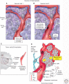
Sources of tumor endothelial cell heterogeneity. (A,B) Tumor endothelial cell phenotypes are determined by the vascular bed of origin (Vascular bed 1 vs. Vascular bed 2), the tumor microenvironment, recruitment and incorporation of bone marrow–derived cells (endothelial progenitor cell, EPC), luminal exposure of tumor cells (A), and genetic instability (B). The different colors represent different tumor endothelial cell phenotypes. The overall differences in color between tumor endothelial cells arising from vascular beds 1 and 2 (A, B, respectively) reflect epigenetically fixed properties. The differences in color between tumor endothelial cells within a given tumor reflect microheterogeneity in the tumor environment. (C) p53 inactivation in tumor cells results in increased hypoxia inducible factor-1α (HIF-1α)-dependent expression of vascular endothelial growth factor (VEGF) and reduced expression of thrombospondin-1 (TSP-1) and collagen (and its antiangiogenic peptides). (D) The tumor microenvironment includes signals on the luminal side, e.g., blood hypoxia, reduced blood flow, and low pH, and on the abluminal side, e.g., mechanical pressure from proliferating tumor cells, tumor-derived paracrine mediators, stromal cell-derived signals, and a disorganized basement membrane. FGF, fibroblast growth factor; VEGF, vascular endothelial growth factor; HGF, hepatocyte growth factor; and EC, endothelial cell. (Figure is from Aird 2009; reprinted, with permission, from the author.)

References

    1. Aird WC 2001. Vascular bed-specific hemostasis: Role of endothelium in sepsis pathogenesis. Crit Care Med 29: S28–S35 - PubMed
    1. Aird WC 2006. Mechanisms of endothelial cell heterogeneity in health and disease. Circ Res 98: 159–162 - PubMed
    1. Aird WC 2007a. Phenotypic heterogeneity of the endothelium: I. Structure, function, and mechanisms. Circ Res 100: 158–173 - PubMed
    1. Aird WC 2007b. Phenotypic heterogeneity of the endothelium: II. Representative vascular beds. Circ Res 100: 174–190 - PubMed
    1. Aird WC 2009. Molecular heterogeneity of tumor endothelium. Cell Tissue Res 335: 271–281 - PubMed

MeSH terms

Substances

LinkOut - more resources