. 2012 Feb 8:9:28.
doi: 10.1186/1742-2094-9-28.

Functional analysis of Pro-inflammatory properties within the cerebrospinal fluid after subarachnoid hemorrhage in vivo and in vitro

Affiliations

Functional analysis of Pro-inflammatory properties within the cerebrospinal fluid after subarachnoid hemorrhage in vivo and in vitro

Ulf C Schneider et al. J Neuroinflammation. .

Abstract

Background: To functionally characterize pro-inflammatory and vasoconstrictive properties of cerebrospinal fluid after aneurysmal subarachnoid hemorrhage (SAH) in vivo and in vitro.

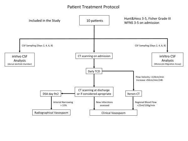
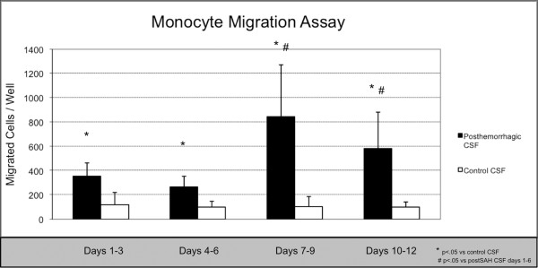
Methods: The cerebrospinal fluid (CSF) of 10 patients suffering from SAH was applied to the transparent skinfold chamber model in male NMRI mice which allows for in vivo analysis of the microcirculatory response to a superfusat. Microvascular diameter changes were quantified and the numbers of rolling and sticking leukocytes were documented using intravital multifluorescence imaging techniques. Furthermore, the pro-inflammatory properties of CSF were assessed in vitro using a monocyte transendothelial migration assay.

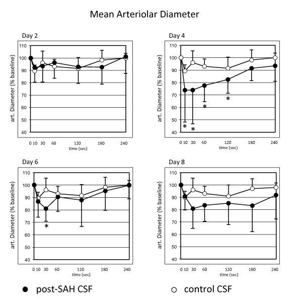
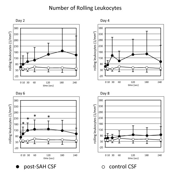
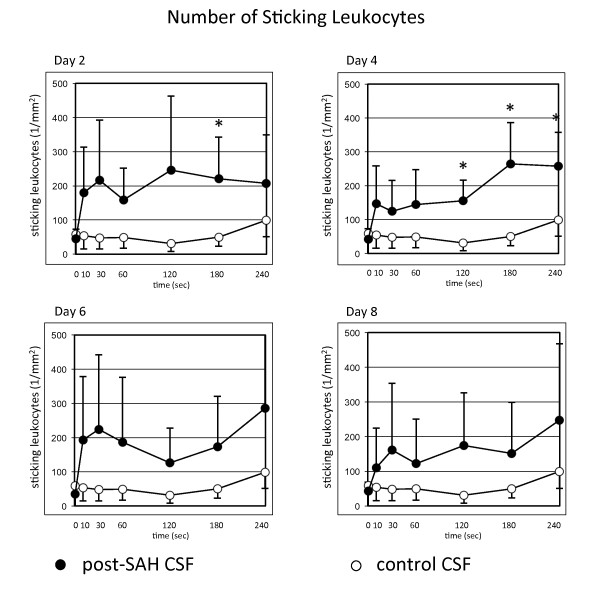
Results: CSF superfusion started to induce significant vasoconstriction on days 4 and 6 after SAH. In parallel, CSF superfusion induced a microvascular leukocyte recruitment, with a significant number of leukocytes rolling (day 6) and sticking (days 2-4) to the endothelium. CSF of patients presenting with cerebral edema induced breakdown of blood vessel integrity in our assay as evidenced by fluorescent marker extravasation. In accordance with leukocyte activation in vivo, significantly higher in vitro monocyte migration rates were found after SAH.

Conclusion: We functionally characterized inflammatory and vasoactive properties of patients' CSF after SAH in vivo and in vitro. This pro-inflammatory milieu in the subarachnoid space might play a pivotal role in the pathophysiology of early and delayed brain injury as well as vasospasm development following SAH.


Figures

Figure 1
Figure 1
Study protocol showing standard patient management, timepoints and decision making of imaging as well as the further processing of post-SAH CSF.
Figure 2
Figure 2
Quantitative analysis of the arteriolar diameter after superfusion of the dorsal skinfold chamber with cerebrospinal fluid (CSF) of patients after severe subarachnoid haemorrhage. Lower diameters are documented after superfusion with post-SAH CSF. Significantly lower diameters are documented on days 4 and 6 after SAH. Values are given as means ± standard deviation. * = p < 0.05 vs. baseline values. N = 10 for post-SAH group (days 2 and 4) and 9 (days 6 and 8), respectively, due to exclusion of one patient's CSF after diagnosis of bacterial meningitis. N = 6 for control group.
Figure 3
Figure 3
Quantitative analysis of rolling leukocytes after superfusion of the dorsal skinfold chamber with post-SAH CSF showing significantly higher numbers of rolling leukocytes on day 6. Values are given as means ± standard deviation. * = p < 0.05 vs. baseline values. N = 10 for post-SAH group (days 2 and 4) and 9 (days 6 and 8), respectively, due to exclusion of one patient's CSF after diagnosis of bacterial meningitis. N = 6 for control group.
Figure 4
Figure 4
Quantitative analysis of sticking leukocytes after superfusion of the dorsal skinfold chamber with post-SAH CSF showing significantly higher numbers of sticking leukocytes on days 2 and 4. Values are given as means ± standard deviation. * = p < 0.05 vs. baseline values. N = 10 for post-SAH group (days 2 and 4) and 9 (days 6 and 8), respectively, due to exclusion of one patient's CSF after diagnosis of bacterial meningitis. N = 6 for control group.
Figure 5
Figure 5
The monocyte transendothelial migration assay showed significantly higher numbers of CD14+ monocyte cells that had migrated through the HUVEC cell layer after stimulation with post-SAH CSF when compared to stimulation with control CSF. * = p < 0.05 vs control CSF # = p < 0.05 vs days 1-3 and days 4-6. N = 10 for post-SAH group (days 1-3) and 9 (days 4-12), respectively, due to exclusion of one patient's CSF after diagnosis of bacterial meningitis. N = 6 for control group.

References

    1. Barth M, Capelle H-H, Weidauer S, Weiss C, Münch E, Thomé C, Luecke T, Schmiedek P, Kasuya H, Vajkoczy P. Effect of nicardipine prolonged-release implants on cerebral vasospasm and clinical outcome after severe aneurysmal subarachnoid hemorrhage: a prospective, randomized, double-blind phase IIa study. Stroke. 2007;38:330–336. doi: 10.1161/01.STR.0000254601.74596.0f. - DOI - PubMed
    1. Vajkoczy P, Meyer B, Weidauer S, Raabe A, Thomé C, Ringel F, Breu V, Schmiedek P. Clazosentan (AXV-034343), a selective endothelin A receptor antagonist, in the prevention of cerebral vasospasm following severe aneurysmal subarachnoid hemorrhage: results of a randomized, double-blind, placebo-controlled, multicenter phase IIa study. J Neurosurg. 2005;103:9–17. doi: 10.3171/jns.2005.103.1.0009. - DOI - PubMed
    1. Hansen-Schwartz J, Vajkoczy P, Macdonald RL, Pluta RM, Zhang JH. Cerebral vasospasm: looking beyond vasoconstriction. Trends Pharmacol Sci. 2007;28:252–256. doi: 10.1016/j.tips.2007.04.002. - DOI - PubMed
    1. Osuka K, Suzuki Y, Tanazawa T, Hattori K, Yamamoto N, Takayasu M, Shibuya M, Yoshida J. Interleukin-6 and development of vasospasm after subarachnoid haemorrhage. Acta Neurochir (Wien) 1998;140:943–951. doi: 10.1007/s007010050197. - DOI - PubMed
    1. Polin RS, Bavbek M, Shaffrey ME, Billups K, Bogaev CA, Kassell NF, Lee KS. Detection of soluble E-selectin, ICAM-1, VCAM-1, and L-selectin in the cerebrospinal fluid of patients after subarachnoid hemorrhage. J Neurosurg. 1998;89:559–567. doi: 10.3171/jns.1998.89.4.0559. - DOI - PubMed

Publication types

MeSH terms

LinkOut - more resources