Review
. 2012;18(9):1173-85.
doi: 10.2174/138161212799436368.

Computational insights for the discovery of non-ATP competitive inhibitors of MAP kinases

Affiliations
Review

Computational insights for the discovery of non-ATP competitive inhibitors of MAP kinases

Michael J Schnieders et al. Curr Pharm Des. 2012.

Abstract

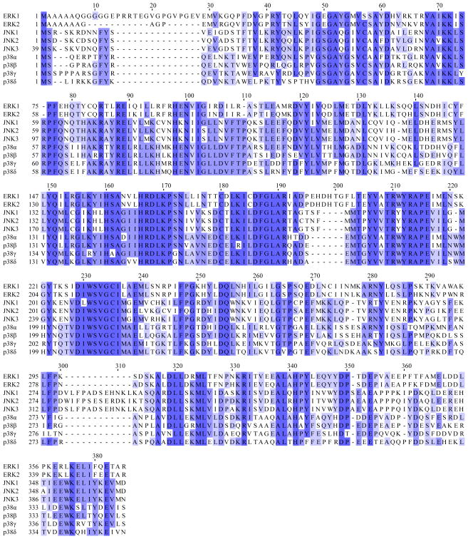
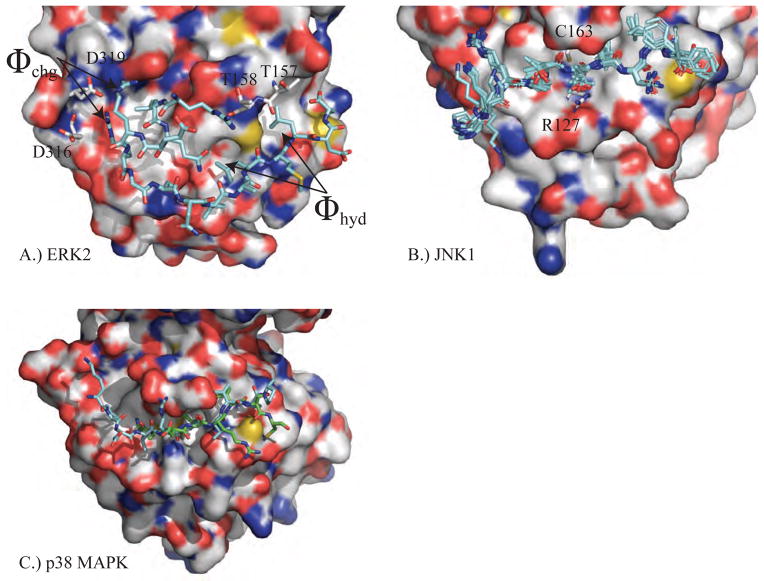
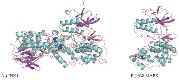
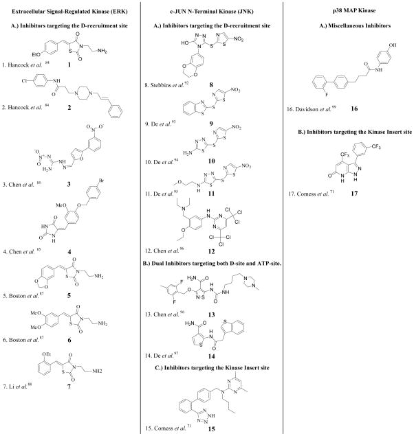
Due to their role in cellular signaling mitogen activated protein (MAP) kinases represent targets of pharmaceutical interest. However, the majority of known MAP kinase inhibitors compete with cellular ATP and target an ATP binding pocket that is highly conserved in the 500 plus representatives of the human protein kinase family. Here we review progress toward the development of non-ATP competitive MAP kinase inhibitors for the extracellular signal regulated kinases (ERK1/2), the c-jun N-terminal kinases (JNK1/2/3) and the p38 MAPKs (α, β, γ, and δ). Special emphasis is placed on the role of computational methods in the drug discovery process for MAP kinases. Topics include recent advances in X-ray crystallography theory that improve the MAP kinase structures essential to structurebased drug discovery, the use of molecular dynamics to understand the conformational heterogeneity of the activation loop and inhibitors discovered by virtual screening. The impact of an advanced polarizable force field such as AMOEBA used in conjunction with sophisticated kinetic and thermodynamic simulation methods is also discussed.


Figures

Figure 1
Figure 1
Shown is a multiple sequence alignment of human ERK1, ERK2, JNK1α2, JNK2α2, JNK3α2 and p38 MAPKα (CSBP2), p38 MAPKβ, p38 MAPKγ and p38 MAPKδ. The shades of blue indicate the degree of conservation for each residue and darker shades correspond to greater conservation. Note that the first 38 n-terminal residues of JNK3α2 and a variable number of c-terminal residues on each MAP kinase are not shown.
Figure 2
Figure 2
A.) JNK1 (PDB ID: 3O17) is shown with each segment of secondary structure labeled. The subscripts for secondary structure elements that are not conserved for all protein kinases refer to loop number. For example, the first β-strand of Loop 0 is β1L0. B.) Ewald refinement of the original experimental data using prior chemical information defined by the polarizable AMOEBA force field improves the quality of the structural model via extension of energetically favorable secondary structure elements and by improving agreement with the measured diffraction data (ie. reducing Rfree from 30.6 to 27.9). The 3 β-strands and 4 α-helices that were extended are labeled. Changes near the DRS-site (Φchg) and FRS-site (αg, activation loop) may impact downstream computational methods such as virtual screening of non-ATP MAP kinase inhibitors.
Figure 3
Figure 3
A.) The inactive structure of ERK2 (PDB ID: 1ERK) shows the unphosphorylated T-X-Y motif (cyan) located on the activation loop (L12) of protein kinases (183T-E-Y185 for ERK1/2). B.) The active structure of ERK2 (PDB ID: 2ERK) has been phosphorylated on both the threonine and tyrosine residues that favor conformational rearrangements stabilized by electrostatic interactions between the negatively charged phosphorylated residues and positively charged arginine residues (grey).
Figure 4
Figure 4
Shown in each panel is a MAP kinase structure complexed with an inhibitor (cyan, spacefill) that targets DFG-in or DFG-out (magenta, ball & stick) and the corresponding conformation of the activation loop (magenta, backbone only). A.) JNK2 in the DFG-in conformation is shown in a complex with type-I inhibitor N-[3-[5-(1H 1,2,4-triazol-3-yl)-1H-indazol-3-yl]phenyl]furan-2-carboxamide (PDB ID 3E7O). B.) Ewald refinement of A orients the water hydrogen-bonding network around the JNK2 inhibitor-binding site. C.) JNK2 in the DFG-out conformation in a complex with type-II inhibitor BIRB-796 (PDB ID: 3NPC). D.) Ewald refinement of C orients the water hydrogen-bonding network around the JNK2 inhibitor-binding site. E.) p38 MAPKα in the DFG-out conformation in a complex with BIRB-796 (PDB ID 1KV2). Ewald refinement was not performed for E because no diffraction data was deposited.
Figure 5
Figure 5
The D-recruitment sites of A.) ERK2 with pepHePTP (PDB ID: 2GPH), B.) JNK1 with pepJIP1 (PDB IDs: 1UKH, 1UKI, 2G01, 2GMX, 2H96, 2NO3) and C.) p38 MAPKα with pepMKK3B (PDB ID: 1LEZ) / pepMEF2A (PDB ID: 1LEW) are shown. Qualitatively, to the right of the conserved C163 residue is a hydrophobic patch that contributes affinity to the interaction of MAP kinases with scaffolding proteins and to the left are interactions with negatively charged aspartate and/or glutamate residues. The superposition of JNK1 structures shows consistency for pepJIP1 residues critical to binding, but significant conformational heterogeneity otherwise.
Figure 6
Figure 6
Shown in A and B are Ewald refinements of two MAP kinase complexes with non-ATP inhibitors that bind at the FRS-site. A.) The structure of the α isoform of JNK1 in complex with inhibitor 46A bound (PDB ID: 3O2M), which exhibits extensive interactions with a second copy of itself within the asymmetric unit of the crystal. B.) A promising structure of p38 MAPKα with inhibitor 3NE bound (PDB ID: 3NEW).
Figure 7
Figure 7
2D structures of non-ATP competitive MAP kinase inhibitors discussed throughout the text.

References

    1. Noble ME, Endicott JA, Johnson LN. Protein kinase inhibitors: insights into drug design from structure. Science. 2004;303:1800–5. - PubMed
    1. Johnson LN. Protein kinase inhibitors: Contributions from structure to clinical compounds. Quarterly reviews of biophysics. 2009;42:1–40. - PubMed
    1. Pearson G, Robinson F, Beers Gibson T, Xu BE, Karandikar M, Berman K, Cobb MH. Mitogen-activated protein (MAP) kinase pathways: Regulation and physiological functions. Endocrine reviews. 2001;22:153–83. - PubMed
    1. Lee S, Warthaka M, Yan C, Kaoud TS, Piserchio A, Ghose R, Ren P, Dalby KN. A model of a MAPK*substrate complex in an active conformation: A computational and experimental approach. PLoS One. 2011;6:e18594. - PMC - PubMed
    1. Thompson JD, Higgins DG, Gibson TJ. CLUSTAL W: improving the sensitivity of progressive multiple sequence alignment through sequence weighting, position-specific gap penalties and weight matrix choice. Nucleic Acids Research. 1994;22:4673–80. - PMC - PubMed

Publication types

MeSH terms

Substances