. 2012 Mar;24(3):288-99.
doi: 10.1111/j.1365-2982.2011.01844.x.

Smooth muscle caldesmon modulates peristalsis in the wild type and non-innervated zebrafish intestine

Affiliations

Smooth muscle caldesmon modulates peristalsis in the wild type and non-innervated zebrafish intestine

J Abrams et al. Neurogastroenterol Motil. 2012 Mar.

Abstract

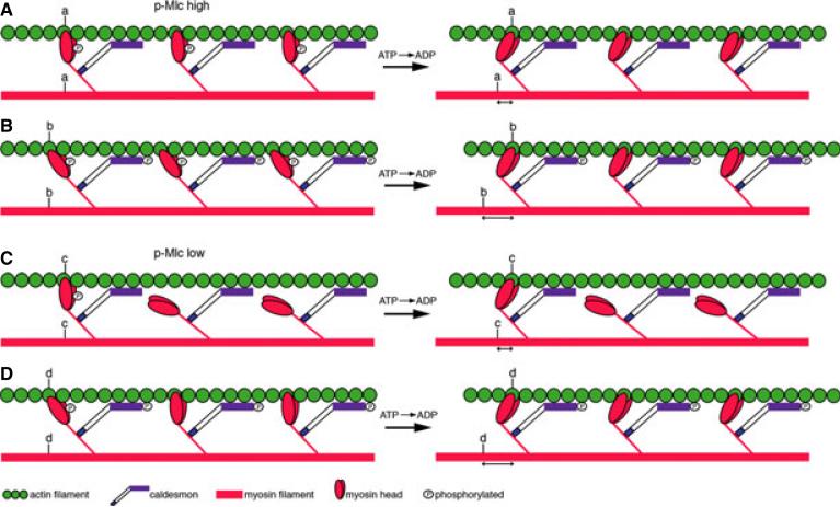
Background: The high molecular weight isoform of the actin-binding protein Caldesmon (h-CaD) regulates smooth muscle contractile function by modulating cross-bridge cycling of myosin heads. The normal inhibitory activity of h-CaD is regulated by the enteric nervous system; however, the role of h-CaD during intestinal peristalsis has never been studied.

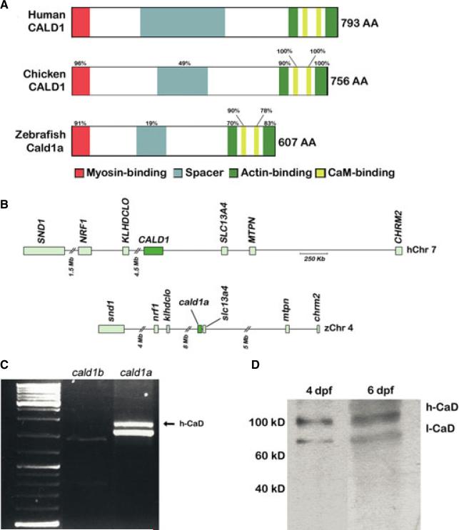
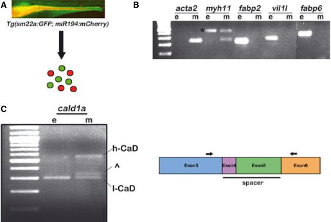
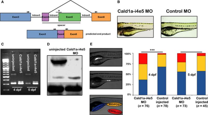
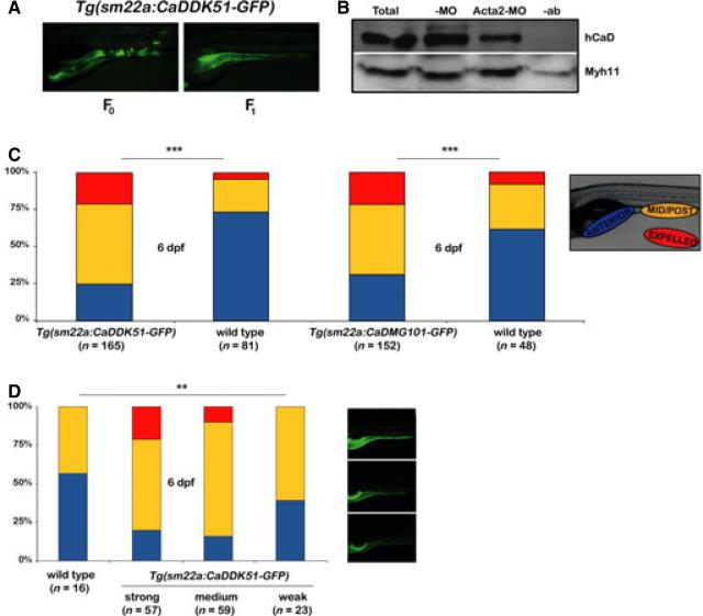
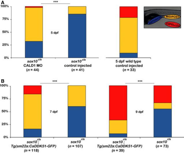
Methods: We identified a zebrafish paralog of the human CALD1 gene that encodes an h-CaD isoform expressed in intestinal smooth muscle. We examined the role of h-CaD during intestinal peristalsis in zebrafish larvae by knocking down the h-CaD protein using an antisense morpholino oligonucleotide. We also developed transgenic zebrafish that express inhibitory peptides derived from the h-CaD myosin and actin-binding domains, and examined their effect on peristalsis in wild-type zebrafish larvae and sox10 (colourless) mutant larvae that lack enteric nerves.

Key results: Genomic analyses identified two zebrafish Caldesmon paralogs. The cald1a ortholog encoded a high molecular weight isoform generated by alternative splicing whose intestinal expression was restricted to smooth muscle. Propulsive intestinal peristalsis was increased in wild-type zebrafish larvae by h-CaD knockdown and by expression of transgenes encoding inhibitory myosin and actin-binding domain peptides. Peristalsis in the non-innervated intestine of sox10 (colourless) larvae was partially restored by h-CaD knockdown and expression of the myosin-binding peptide.

Conclusions & inferences: Disruption of the normal inhibitory function of h-CaD enhances intestinal peristalsis in both wild-type zebrafish larvae and mutant larvae that lack enteric nerves, thus confirming a physiologic role for regulation of smooth muscle contraction at the actin filament.


Figures

Figure 1
Figure 1
Simplified model of h-CaD function. Smooth muscle contraction depicted by sliding of actin (green) on myosin (red) filaments, following ATP hydrolysis (derived from previously described model). (A) In smooth muscle with high levels of phospho-Mlc (p-Mlc), non-phosphorylated h-CaD restricts binding of myosin heads to the actin filament such that force generation is inhibited. (B) When h-CaD is phosphorylated, the myosin heads are able to bind the actin filament in a manner that enhances actomyosin interaction (b). This leads to increased force generation (distance b > distance a). (C) In smooth muscle with low levels of p-Mlc, non-phosphorylated h-CaD prevents binding of non-phosphorylated myosin heads to actin. (D) phospho-h-CaD promotes binding of the non-phosphorylated myosin heads to actin, thereby enhancing contraction; (distance d > distance c). Distances a, b and c, d are not drawn to scale.
Figure 2
Figure 2
Zebrafish smooth muscle Caldesmon (h-CaD). (A) Schematic representation of the human, chicken and zebrafish h-CaD orthologs. Conserved protein domains and percent amino acid homology are indicated. (B) Conserved syntenic relationships surrounding the human CALD1 locus on chromosome 7 and the zebrafish cald1a locus on chromosome 4. (C) RT-PCR showing amplification of the full length cDNA corresponding to the high and low molecular weight zebrafish cald1a isoforms from intestinal cDNA. A correctly sized transcript for the predicted low molecular weight cald1b isoform is also detected. (D) Western blot showing intestinal levels of CaD isoforms. Molecular weight standards indicated. Mb, megabase; Kb, kilobase; dpf, days post fertilization.
Figure 3
Figure 3
Expression of the zebrafish h-CaD ortholog in intestinal smooth muscle. (A) Scheme to isolate intestinal smooth muscle (green) and epithelial (red) cells from Tg(sm22a:GFP; miR194:mCherry) larvae. Fluorescent image of the intestine of a 5 dpf bigenic larva is shown. Smooth muscle of the anterior intestine is not in plane of focus. (B) RT-PCR amplification showing expression of intestinal smooth muscle and epithelial markers in sorted cells. The myh11 primers amplify a band (*) from contaminating epithelial and smooth muscle genomic DNA (confirmed by sequencing). (C) Expression of the high molecular weight cald1a transcript is restricted to smooth muscle. In some experiments, an additional band was amplified from smooth muscle that migrated near the low molecular weight cald1 transcript (^); however, this was detected in only a minority of experiments. Schematic indicates the location of PCR primers (arrows) in exon 3 and exon 6 that were used to amplify the cald1a isoforms. e, epithelial; m, smooth muscle.
Figure 4
Figure 4
h-CaD deficiency enhances intestinal peristalsis in zebrafish larvae. (A) Schematic depicting isoform specific targeting of the high molecular weight cald1a transcript by a splice blocking morpholino (Cald1a-i4e5 MO). The exon 5 splice acceptor is targeted (*). (B) Normal morphology of 4 dpf larvae injected with Cald1a-i4e5 MO and control morpholinos. (C) RT-PCR of intestinal cDNA from Cald1a-i4e5 MO larvae using exon 3 and exon 6 primers (arrows in panel 3A) shows markedly reduced expression of the high molecular weight cald1a transcript. (D) Western blot using intestinal protein from Cald1a-i4e5 MO larvae shows reduced levels of h-CaD with slightly increased l-CaD levels. Increased protein corresponding to l-CaD likely reflects additional protein translated from modified transcript only 12 amino acids larger than l-CaD. (E) Images of live 5 dpf larvae that ingested fluorescent microspheres located in anterior and mid-posterior intestine, respectively. Color scheme in bar graph depicts bead location indicated in lateral image of larva (lower panel). Cald1a-i4e5 MO larvae show increased propulsive peristalsis at 4dpf (chi-squared test, ***P < 001). 5dpf P-value = 0.08, likely due to transient effect of morpholino knockdown.
Figure 5
Figure 5
Expression of peptides blocking h-CaD interaction at myosin and actin filaments increases intestinal propulsive peristalsis. (A) Lateral images of live 5 dpf F0 and F1 Tg(sm22a: CaDDK51-GFP) larvae showing mosaic and widespread smooth muscle GFP expression, respectively. (B) IP-Western blot confirms that CaDDK51 myosin-binding domain peptide blocks interaction of h-CaD with Myh11. Total intestinal protein was IP'ed with Myh11 antibody and blotted as indicated. (C) Bar graph shows increased propulsive peristalsis in unsorted Tg(sm22a: CaDDK51 -GFP) larvae and Tg (sm22a: CaDMG101 -GFP) larvae, in which peptides block h-CaD interaction with myosin and actin, respectively (chi-squared test, ***P < 001). (D) Enhanced peristalsis in Tg (sm22a: CaDDK51 -GFP) larvae correlates with the level of transgene expression (determined by GFP fluorescence). Images show larvae with strong, medium and weak GFP expression (Fisher's exact test, **P < 05). -MO – control larvae; Acta2-MO – larvae injected with smooth muscle actin morpholino; -ab – control IP without antibody; Myh11 – smooth muscle myosin.
Figure 6
Figure 6
Intestinal peristalsis is increased in h-CaD deficient colourless larvae. (A) Bar graph shows partial rescue of intestinal peristalsis in h-CaD deficient 5 dpf cls larvae. (B) Tg (sm22a: CaDDK51 -GFP) cls larvae show partial rescue of propulsive peristalsis at 7 dpf and 9 dpf (chi-squared test, ***P < 001).

References

    1. Kamm KE, Stull JT. The function of myosin and myosin light chain kinase phosphorylation in smooth muscle. Annu Rev Pharmacol Toxicol. 1985;25:593–620. - PubMed
    1. Murphy RA. Contraction in smooth muscle cells. Annu Rev Physiol. 1989;51:275–83. - PubMed
    1. Siegman MJ, Butler TM, Mooers SU, Michalek A. Ca2+ can affect Vmax without changes in myosin light chain phosphorylation in smooth muscle. Pflugers Arch. 1984;401:385–90. - PubMed
    1. Gerthoffer WT. Dissociation of myosin phosphorylation and active tension during muscarinic stimulation of tracheal smooth muscle. J Pharmacol Exp Ther. 1987;240:8–15. - PubMed
    1. Haeberle JR, Hott JW, Hathaway DR. Regulation of isometric force and isotonic shortening velocity by phosphorylation of the 20,000 dalton myosin light chain of rat uterine smooth muscle. Pflugers Arch. 1985;403:215–9. - PubMed

Publication types