. 2012 Feb 8:9:4.
doi: 10.1186/2045-8118-9-4.

Evaluation of blood-brain barrier transport and CNS drug metabolism in diseased and control brain after intravenous L-DOPA in a unilateral rat model of Parkinson's disease

Affiliations

Evaluation of blood-brain barrier transport and CNS drug metabolism in diseased and control brain after intravenous L-DOPA in a unilateral rat model of Parkinson's disease

Paulien Gm Ravenstijn et al. Fluids Barriers CNS. .

Abstract

Background: Changes in blood-brain barrier (BBB) functionality have been implicated in Parkinson's disease. This study aimed to investigate BBB transport of L-DOPA transport in conjunction with its intra-brain conversion, in both control and diseased cerebral hemispheres in the unilateral rat rotenone model of Parkinson's disease.

Methods: In Lewis rats, at 14 days after unilateral infusion of rotenone into the medial forebrain bundle, L-DOPA was administered intravenously (10, 25 or 50 mg/kg). Serial blood samples and brain striatal microdialysates were analysed for L-DOPA, and the dopamine metabolites DOPAC and HVA. Ex-vivo brain tissue was analyzed for changes in tyrosine hydroxylase staining as a biomarker for Parkinson's disease severity. Data were analysed by population pharmacokinetic analysis (NONMEM) to compare BBB transport of L-DOPA in conjunction with the conversion of L-DOPA into DOPAC and HVA, in control and diseased cerebral hemisphere.

Results: Plasma pharmacokinetics of L-DOPA could be described by a 3-compartmental model. In rotenone responders (71%), no difference in L-DOPA BBB transport was found between diseased and control cerebral hemisphere. However, in the diseased compared with the control side, basal microdialysate levels of DOPAC and HVA were substantially lower, whereas following L-DOPA administration their elimination rates were higher.

Conclusions: Parkinson's disease-like pathology, indicated by a huge reduction of tyrosine hydroxylase as well as by substantially reduced levels and higher elimination rates of DOPAC and HVA, does not result in changes in BBB transport of L-DOPA. Taking the results of this study and that of previous ones, it can be concluded that changes in BBB functionality are not a specific characteristic of Parkinson's disease, and cannot account for the decreased benefit of L-DOPA at later stages of Parkinson's disease.


Figures

Figure 1
Figure 1
The population pharmacokinetic model for L-DOPA, DOPAC and HVA comprising of three compartments (1-3) describing the pharmacokinetics of L-DOPA in plasma, two compartments (4 and 5) describing the pharmacokinetics of L-DOPA in brainECF, one for the control cerebral hemisphere and one for the rotenone-treated responder cerebral hemisphere, two compartments (6 and 8) describing the kinetics of DOPAC in brainECF, one for the control cerebral hemisphere and one for the rotenone-treated responder cerebral hemisphere and two compartments (7 and 9) describing the kinetics of HVA in brainECF, one for the control cerebral hemisphere and one for the rotenone-treated responder cerebral hemisphere. (V = volume of distribution, Q = inter-compartmental clearance, k = elimination rate constant, Kin = endogenous formation rate constant of L-DOPA.).
Figure 2
Figure 2
L-DOPA concentration-time profiles in plasma, obtained after a 20-min intravenous infusion in Lewis rats. Depicted are the observed concentrations (dots) and individual predictions (solid lines), separated by L-DOPA dose (in total 13 rats: 10 mg/kg, n = 4; 25 mg/kg, n = 4; 50 mg/kg, n = 5).
Figure 3
Figure 3
L-DOPA concentration-time profiles in brainECF in the control cerebral hemisphere (○; in total 12 rats: 10 mg/kg, n = 4; 25 mg/kg, n = 3; 50 mg/kg, n = 5) and in the rotenone-treated responder cerebral hemisphere (•; in total 7 rats: 10 mg/kg, n = 1; 25 mg/kg, n = 2; 50 mg/kg, n = 4), obtained after a 20-min intravenous infusion in Lewis rats.
Figure 4
Figure 4
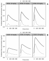
Population predicted concentrations of DOPAC (panel A) and HVA (panel B) in the control cerebral hemisphere (-) and rotenone-treated responder cerebral hemisphere in responders (---) according to the model as described in Figure 1, at three doses of L-DOPA.
Figure 5
Figure 5
Individual average baseline concentrations (ng/ml) of L-DOPA (A), DOPAC (B) and HVA (C) as a function of the decrease in TH staining (%).

References

    1. Elsworth JD, Roth RH. Dopamine synthesis, uptake, metabolism, and receptors: relevance to gene therapy of Parkinson's disease. Exp Neurol. 1997;144:4–9. doi: 10.1006/exnr.1996.6379. - DOI - PubMed
    1. Westerink BH. Sequence and significance of dopamine metabolism in the rat brain. Neurochem Int. 1985;7:221–227. doi: 10.1016/0197-0186(85)90108-1. - DOI - PubMed
    1. Männistö PT, Ulmanen I, Lundström K, Taskinen J, Tenhunen J, Tilgmann C, Kaakkola S. Characteristics of catechol O-methyl-transferase (COMT) and properties of selective COMT inhibitors. Prog Drug Res. 1992;39:291–350. - PubMed
    1. Dauer W, Przedborski S. Parkinson's disease: mechanisms and models. Neuron. 2003;39:889–909. doi: 10.1016/S0896-6273(03)00568-3. - DOI - PubMed
    1. Deleu D, Northway MG, Hanssens Y. Clinical pharmacokinetic and pharmacodynamic properties of drugs used in the treatment of Parkinson's disease. Clin Pharmacokinet. 2002;41:261–309. doi: 10.2165/00003088-200241040-00003. - DOI - PubMed

LinkOut - more resources