. 2012 Mar;71(3):198-210.
doi: 10.1097/NEN.0b013e3182482590.

Unmyelinated axons show selective rostrocaudal pathology in the corpus callosum after traumatic brain injury

Affiliations

Unmyelinated axons show selective rostrocaudal pathology in the corpus callosum after traumatic brain injury

Thomas M Reeves et al. J Neuropathol Exp Neurol. 2012 Mar.

Abstract

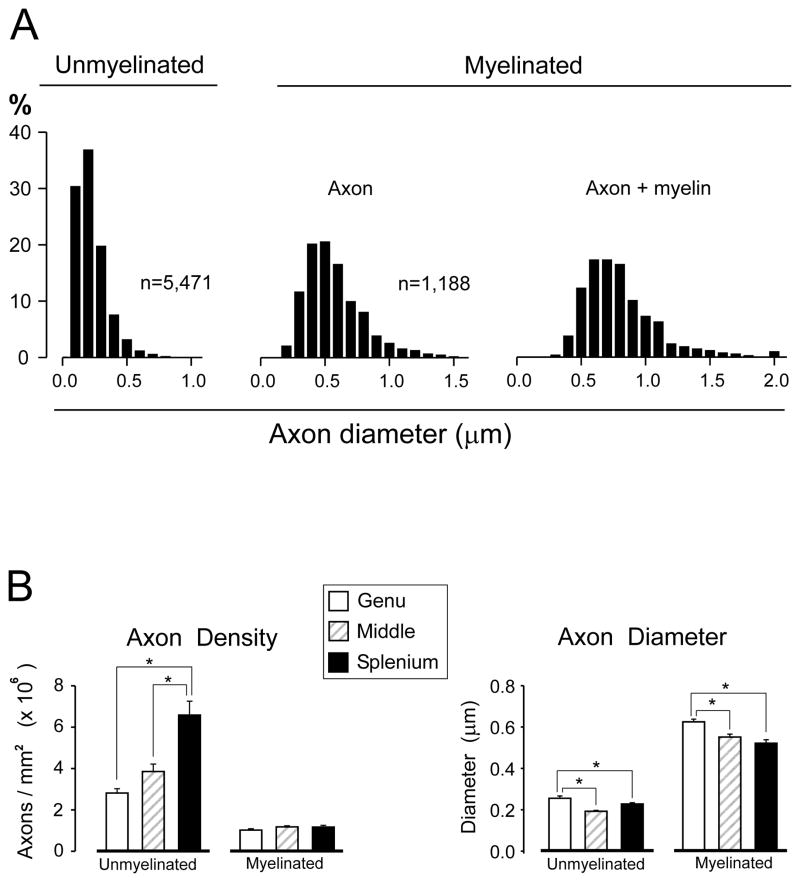
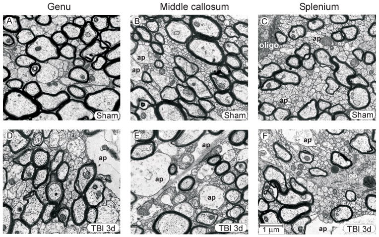
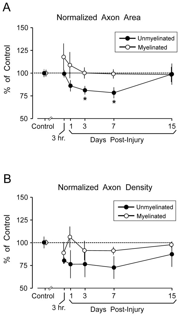
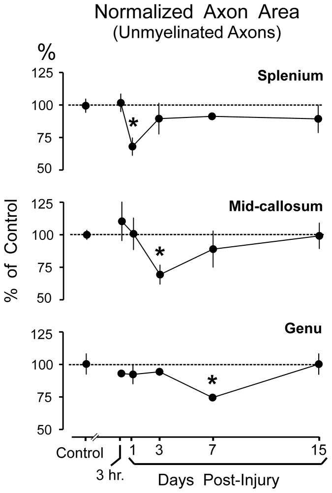
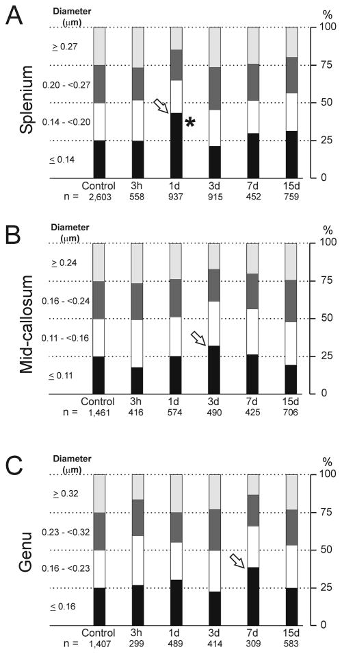
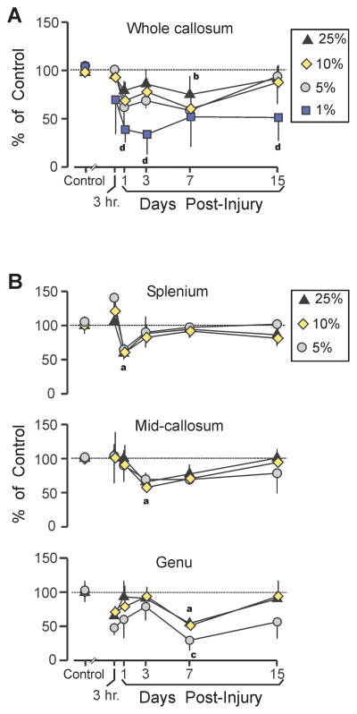
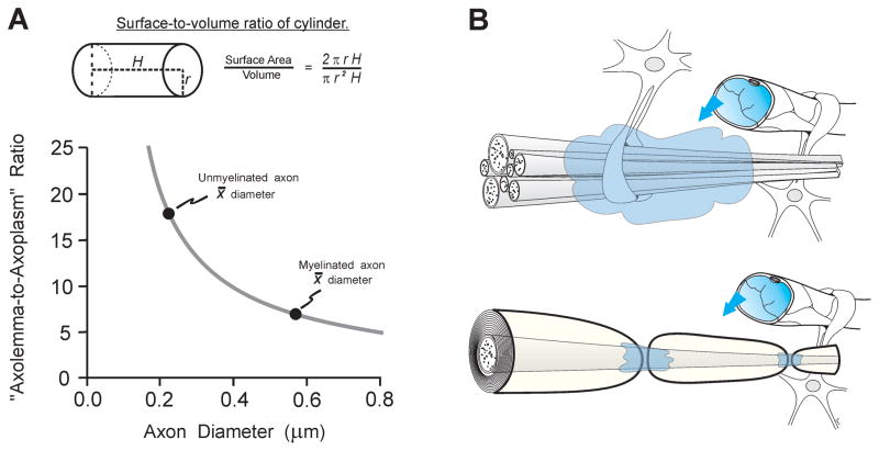
Axonal injury is consistently observed after traumatic brain injury (TBI). Prior research has extensively characterized the post-TBI response in myelinated axons. Despite evidence that unmyelinated axons comprise a numerical majority of cerebral axons, pathologic changes in unmyelinated axons after TBI have not been systematically studied. To identify morphologic correlates of functional impairment of unmyelinated fibers after TBI, we assessed ultrastructural changes in corpus callosum axons. Adult rats received moderate fluid percussion TBI, which produced diffuse injury with no contusion. Cross-sectional areas of 13,797 unmyelinated and 3,278 intact myelinated axons were stereologically measured at survival intervals from 3 hours to 15 days after injury. The mean caliber of unmyelinated axons was significantly reduced at 3 to 7 days and recovered by 15 days, but the time course of this shrinkage varied among the genu, mid callosum, and splenium. Relatively large unmyelinated axons seemed to be particularly vulnerable. Injury-induced decreases in unmyelinated fiber density were also observed, but they were more variable than caliber reductions. By contrast, no significant morphometric changes were observed in myelinated axons. The finding of a preferential vulnerability in unmyelinated axons has implications for current concepts of axonal responses after TBI and for development of specifically targeted therapies.


Figures

Figure 1
Figure 1
(A) Midsagittal diagram of rat brain, showing sampling scheme covering the dorsal-to-ventral extent of the corpus callosum at genu [G], mid-callosum [M], and splenium [S]. Shaded region shows width and location of craniotomy for midline fluid percussion injury. (B) Example of an unbiased stereological counting frame. Shaded profiles [d] show example unmyelinated axon profiles. Representative myelinated profile shows 3 contours that were digitally traced for area measurements: a, outer myelin border; b, inner myelin border; c, axolemma.
Figure 2
Figure 2
Characterization of axon populations in sham lesion control rats. (A) Distributions of axon diameters for myelinated and unmyelinated axons. Mean diameters and distribution shapes matched prior reports from naïve control rodents (see text), suggesting that the sham lesion procedure did not impact these parameters. (B) Separate analyses at callosal regions revealed a rostral-to-caudal (genu-to-splenium) increase in the density of unmyelinated axons, but no gradient for myelinated axons (left panel). Mean axon diameter was significantly greater in the genu than in either posterior region (right panel). *p < 0.05.
Figure 3
Figure 3
Representative micrographs, showing genu, mid-callosum, and splenium regions in sham injured control rats (A–C) and during the post-injury period when fluid percussion injury-induced change in the unmyelinated axons was well developed (3 days post-injury) (D–F). Even though quantitative analyses revealed significant morphometric changes to axonal dimensions at 3 days post-injury, the general architecture of injured tissue was similar to that in sham control tissue, although in some injured rats astrocyte processes were larger and more frequently encountered. The rostral-to-caudal increase in unmyelinated axon density, noted in sham control rats, is preserved in the post-injury samples. Abbreviations: ap, astrocyte process; oligo, oligodendrocyte cell body; op, oligodendrocyte process. Calibration bar in F applies to all panels.
Figure 4
Figure 4
Effect of moderate fluid percussion injury (FPI) on cross-sectional area of axons and axon density. (A) Mean cross-sectional area of axons, averaged across all callosal regions (genu, mid-callosum, and splenium). Results are normalized to the mean axonal area measured in sham injured control rats. Time-dependent axonal shrinkage of unmyelinated fibers was significant (*p < 0.05) at 3 days and 7 days post-injury, but recovered to control levels by 15 days. FPI did not significantly alter cross-sectional areas of myelinated axons. (B) Mean normalized axon density, averaged across all callosal regions. FPI effects on axonal density were more variable, and reductions in unmyelinated fiber density did not reach significance at any single survival interval. FPI did not significantly alter density of myelinated axons. Data points in A and B are slightly offset to avoid overlap of error bars.
Figure 5
Figure 5
Representative axonal profiles that failed to meet the operational definition of ‘intact’ in the splenium at 3 days post-injury. (A) Isolated unmyelinated axons exhibiting membrane discontinuities (arrowhead) and membranous folding apposed to aberrant extracellular spaces (arrow). (B) Clusters of unmyelinated fibers lacking distinct membranes (arrows), along with a myelinated axon (asterisk) with cytoplasmic abnormalities. (C) Example of vulnerability of relatively large unmyelinated axons (arrows) juxtaposed to intact small unmyelinated axons. All calibration as in panel C.
Figure 6
Figure 6
Effect of moderate fluid percussion injury (FPI) on cross-sectional area of unmyelinated axons in the splenium, middle, and genu regions of the corpus callosum. Results are normalized to the mean axonal area measured in sham injured control rats. FPI produced a caudal-to-rostral sequence of significant axonal shrinkage expressed at 1 day in the splenium, 3 days in the mid-callosum, and 7 days in the genu. Each region showed a recovery to sham control levels following the shrinkage. *p < 0.05.
Figure 7
Figure 7
Fluid percussion injury (FPI) effects on quartile distribution of unmyelinated axon diameters. Quartile values were calculated for the diameters of unmyelinated axons in sham injured control rats (first column), for splenium (A), mid-callosum (B), and genu (C). Columns track post-injury changes over time in the percentage of axons falling in each range. Decreases in the largest quartile coincided with increases in bottom quartile at 1 day (splenium), 3 days (mid-callosum) and 7 day (genu) (arrows), a temporal pattern matching maximum reductions in unmyelinated fiber cross-section areas. Post-injury increases in the lowest quartile were only significant for the splenium (*p < 0.05).
Figure 8
Figure 8
Effects of traumatic brain injury (TBI) on density of largest intact unmyelinated axons. (A) Following fluid percussion injury (FPI), the mean density of the largest 25%, 10%, and 5% diameter intact fibers, combined across all callosal regions, was below sham control levels (significant for top 10% fibers at 7 day), but showed a trend toward a recovery by 15 days. In contrast, the density of the largest 1% fiber category was significantly decreased at 1 day, 3 days, and 15 days. (B) Density changes in largest unmyelinated fibers, showed a caudal-to-rostral sequence spanning 1 day to 7 days, similar to post-injury diameter changes (Fig. 6). The largest 5% fiber category was significantly below control levels only in the genu at 7 days. Sham vs. TBI comparisons (all p < 0.05): largest 25% ‘a’, 10% ‘b’, 5% ‘c’, 1% ‘d’.
Figure 9
Figure 9
Morphological properties of unmyelinated axons may place them at elevated risk to traumatic brain injury (TBI). (A) Modeling axons as uniform cylinders of arbitrary length, a decrease in diameter corresponds to an increase in surface-to-volume ratio (upper panel). This increased ratio of axolemma area to axoplasm volume probably constitutes a risk to membrane-targeting pathogenetic mechanisms. Mean diameters for unmyelinated and myelinated axons measured in the present study are plotted on the curve-relating diameter to the surface-to-volume ratio (lower panel). The mean diameter of unmyelinated axons is about 60% smaller than the mean myelinated diameter, but this corresponds to an approximate 160% increase in the surface-to-volume ratio. (B) Diagram of unmyelinated axons and a myelinated axon in relation to non-neuronal elements (blood vessels, astrocytes). Unmyelinated axolemma is more exposed than myelinated axolemma to destructive extracellular influences (shading) arising locally (e.g. ionic imbalances and activated proteases) or from blood-borne factors penetrating through a compromised blood-brain barrier (arrows). These structural differences likely contribute to a greater vulnerability of unmyelinated axons in some injury conditions.

References

    1. Rutland-Brown W, Langlois JA, Thomas KE, et al. Incidence of traumatic brain injury in the United States, 2003. J Head Trauma Rehabil. 2006;21:544–8. - PubMed
    1. Skandsen T, Kvistad KA, Solheim O, et al. Prevalence and impact of diffuse axonal injury in patients with moderate and severe head injury: a cohort study of early magnetic resonance imaging findings and 1-year outcome. J Neurosurg. 2010;113:556–63. - PubMed
    1. Meythaler JM, Peduzzi JD, Eleftheriou E, et al. Current concepts: diffuse axonal injury-associated traumatic brain injury. Arch Phys Med Rehabil. 2001;82:1461–71. - PubMed
    1. Strich SJ. Shearing of nerve fibers as a cause of brain damage due to head injury: a pathological study of twenty cases. Lancet. 1961;2:443–8.
    1. Maxwell WL, Povlishock JT, Graham DL. A mechanistic analysis of nondisruptive axonal injury: a review. J Neurotrauma. 1997;14:419–40. - PubMed

Publication types