. 2012 Apr;86(8):4358-69.
doi: 10.1128/JVI.07107-11. Epub 2012 Feb 8.

Immunodominant "asymptomatic" herpes simplex virus 1 and 2 protein antigens identified by probing whole-ORFome microarrays with serum antibodies from seropositive asymptomatic versus symptomatic individuals

Affiliations

Immunodominant "asymptomatic" herpes simplex virus 1 and 2 protein antigens identified by probing whole-ORFome microarrays with serum antibodies from seropositive asymptomatic versus symptomatic individuals

Gargi Dasgupta et al. J Virol. 2012 Apr.

Abstract

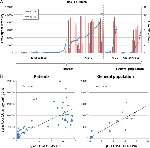
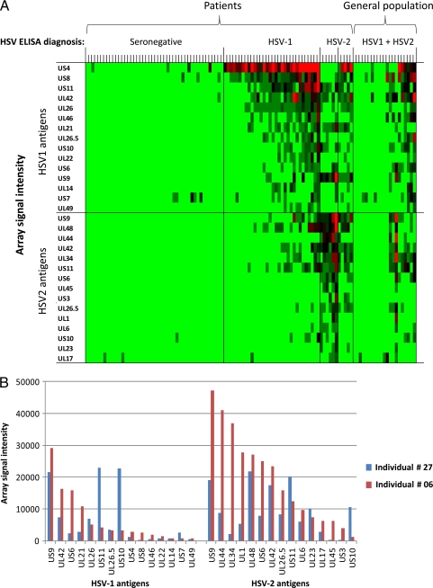
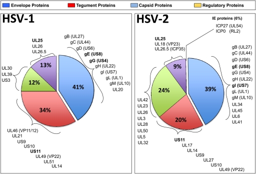
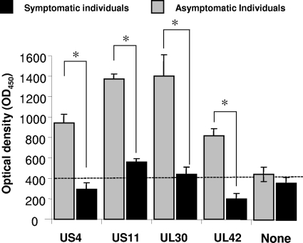
Herpes simplex virus 1 (HSV-1) and HSV-2 are medically significant pathogens. The development of an effective HSV vaccine remains a global public health priority. HSV-1 and HSV-2 immunodominant "asymptomatic" antigens (ID-A-Ags), which are strongly recognized by B and T cells from seropositive healthy asymptomatic individuals, may be critical to be included in an effective immunotherapeutic HSV vaccine. In contrast, immunodominant "symptomatic" antigens (ID-S-Ags) may exacerbate herpetic disease and therefore must be excluded from any HSV vaccine. In the present study, proteome microarrays of 88 HSV-1 and 84 HSV-2 open reading frames(ORFs) (ORFomes) were constructed and probed with sera from 32 HSV-1-, 6 HSV-2-, and 5 HSV-1/HSV-2-seropositive individuals and 47 seronegative healthy individuals (negative controls). The proteins detected in both HSV-1 and HSV-2 proteome microarrays were further classified according to their recognition by sera from HSV-seropositive clinically defined symptomatic (n = 10) and asymptomatic (n = 10) individuals. We found that (i) serum antibodies recognized an average of 6 ORFs per seropositive individual; (ii) the antibody responses to HSV antigens were diverse among HSV-1- and HSV-2-seropositive individuals; (iii) panels of 21 and 30 immunodominant antigens (ID-Ags) were identified from the HSV-1 and HSV-2 ORFomes, respectively, as being highly and frequently recognized by serum antibodies from seropositive individuals; and (iv) interestingly, four HSV-1 and HSV-2 cross-reactive asymptomatic ID-A-Ags, US4, US11, UL30, and UL42, were strongly and frequently recognized by sera from 10 of 10 asymptomatic patients but not by sera from 10 of 10 symptomatic patients (P < 0.001). In contrast, sera from symptomatic patients preferentially recognized the US10 ID-S-Ag (P < 0.001). We have identified previously unreported immunodominant HSV antigens, among which were 4 ID-A-Ags and 1 ID-S-Ag. These newly identified ID-A-Ags could lead to the development of an efficient "asymptomatic" vaccine against ocular, orofacial, and genital herpes.


Figures

Fig 1
Fig 1
Validation of the microarray. (A) The protein microarray signals for HSV-1 US4/gG were compared to OD450 readings by a FocuSelect ELISA using sera from 90 patients and 21 randomly selected general population controls, The patients were grouped according to serodiagnosis based on the commercial ELISA results and ranked within each group by the signal to HSV-1 US4/gG on the array. (B) Linear regression analysis between the sum of the top 10 HSV-1 antigens recognized on the array by HSV-1-seropositive individuals and ODs for patient sera (left) and population controls (right) determined by a FocuSelect ELISA.
Fig 2
Fig 2
Intensities of antibody responses from HSV-1- and HSV-2-seropositive individuals against HSV-1 and HSV-2 antigens. (A) Heat map overview of broadly targeted HSV-1 and HSV-2 immunodominant antigens determined by the intensities of antibody responses. Columns correspond to sera used to probe the array, and rows are arrayed antigens. Sera were serotyped by using FocuSelect 1 and 2 IgG ELISAs (Focus Diagnostics), as shown at the top, and were used as the reference for sample categorizations. The patient sera were thus classified into seronegative (n = 47), HSV-1-seropositive-only (n = 32), HSV-2-seropositive-only (n = 6), and HSV-1- and HSV-2-seropositive (n = 5) groups. For comparison, sera from the general population were probed (n = 21). Only those antigens that were reactive against sera from the HSV-1- or HSV-2-seropositive populations are shown. An antigen was defined as reactive when the average signal intensity for a donor population was more than the mean plus 2 SD of the control spots consisting of IVTT reaction mixtures lacking a DNA template (C plus 2 SD). The HSV-1 antigens are ranked by descending average signal intensity of the HSV-1-seropositive population, and the HSV-2 antigens are similarly ranked by the HSV-2-positive population. In each case, only the top 15 antigens are shown. The sera are also ranked from left to right within each group by the increasing sum of the signals. The heat map was generated from log-normalized data that were retransformed to approximate raw values, and the signal was converted into a color (red, high; green, low). (B) Representative data for serum antibodies from two HSV-2-seropositive individuals recognizing protein microarrays derived from HSV-2 ORFs. IgG antibodies from seropositive individuals 6 and 27 reacted strongly to 7 and 11 ORFs, respectively.
Fig 3
Fig 3
Pie chart summarizing the proportions of HSV-1 and HSV-2 envelope glycoproteins, tegument proteins, capsid proteins, and regulatory proteins recognized by serum antibodies from HSV-1- and/or HSV-2-seropositive individuals. Note that HSV-2 US4/gG was not present on the array.
Fig 4
Fig 4
Intensities of antibody responses against four immunodominant HSV-1 antigens from symptomatic and asymptomatic patients. The average intensity of recognition of RTS expressing US4, US11, UL30, and UL42 proteins by serum antibodies from HSV-seropositive symptomatic (n = 10) and asymptomatic patients (n = 10) was determined by an ELISA. The optical density (OD) at 450 nm determined by an ELISA is shown for each serum sample. The P value was calculated by t tests. Note the difference in serum antibody responses induced by US11 and UL30 in asymptomatic versus symptomatic patients. The antibody recognition of four antigens (US4, US11, UL30, and UL42) was different by >2-fold between serum samples from symptomatic and asymptomatic subjects.

References

    1. Anderson KS, et al. 2008. Application of protein microarrays for multiplexed detection of antibodies to tumor antigens in breast cancer. J. Proteome Res. 7:1490–1499 - PMC - PubMed
    1. Anderson KS, et al. 2011. Protein microarray signature of autoantibody biomarkers for the early detection of breast cancer. J. Proteome Res. 10:85–96 - PMC - PubMed
    1. Barbour AG, et al. 2008. A genome-wide proteome array reveals a limited set of immunogens in natural infections of humans and white-footed mice with Borrelia burgdorferi. Infect. Immun. 76:3374–3389 - PMC - PubMed
    1. Belshe PB, et al. 2012. Efficacy results of a trial of a herpes simplex vaccine. N. Engl. J. Med. 366:34–43 - PMC - PubMed
    1. BenMohamed L, et al. 2003. Identification of novel immunodominant CD4+ Th1-type T-cell peptide epitopes from herpes simplex virus glycoprotein D that confer protective immunity. J. Virol. 77:9463–9473 - PMC - PubMed

Publication types

MeSH terms

LinkOut - more resources