

doi: 10.4161/onci.1.1.17824.
PD-L1 co-stimulation, ligand-induced TCR down-modulation and anti-tumor immunotherapy
Affiliations
- PMID: 22318430
- PMCID: PMC3272426
- DOI: 10.4161/onci.1.1.17824
Item in Clipboard
PD-L1 co-stimulation, ligand-induced TCR down-modulation and anti-tumor immunotherapy
Oncoimmunology.
.
Abstract
PD-1 engagement on the surface of effector T cells strongly suppresses their cytotoxic function, which constitutes a major obstacle for T cell-mediated anti-tumor activities. Surprisingly, PD-1 is strongly upregulated in T cells, engaging its ligand PD-L1 during antigen presentation. However, our recent published data may provide an explanation for this apparent contradiction.
Figures
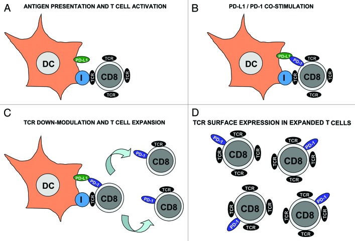
Regulatory role of PD-1 during T cell activation after antigen presentation. (A) Dendritic cells (DC, as indicated) present antigenic peptide associated with MHC class I (encircled “I”) to specific CD8 T cells. DCs also express PD-L1 (green ovoid) on their surface. CD8 T cells associate with DCs through their TCR (black ovoid) as shown in the figure. These CD8 T cells are TCRhigh and do not express PD-1. (B) After antigen recognition, T cells express PD-1 on their surface (blue ovoid), where it engages with PD-L1 on the DC surface. Binding of PD-1 reduces TCR signal transduction and (C) down-modulates TCR levels in CD8 T cells. Activated CD8 T cells proliferate as indicated by arrows. Please note that these proliferating CD8 T cells are TCRlow PD-1+. It is tempting to speculate that low TCR and high PD-1 may inhibit autoreactive cytotoxicity while effector CD8 T cells are undergoing expansion. (D) Expanded effector CD8 T cells gradually recover their TCR surface expression while keeping PD-1 expression. It could be argued that this increase in TCR levels may “arm” cytotoxic T cells against their targets.
References
Grants and funding
LinkOut - more resources
Full Text Sources
Other Literature Sources
Research Materials