Randomized Controlled Trial
. 2012 Jul;91(7):1097-105.
doi: 10.1007/s00277-011-1404-z. Epub 2012 Feb 10.

N-acetylcysteine reduces oxidative stress in sickle cell patients

Affiliations
Randomized Controlled Trial

N-acetylcysteine reduces oxidative stress in sickle cell patients

Erfan Nur et al. Ann Hematol. 2012 Jul.

Abstract

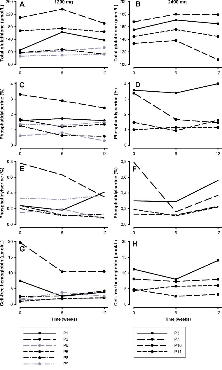
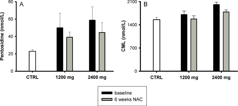
Oxidative stress is of importance in the pathophysiology of sickle cell disease (SCD). In this open label randomized pilot study the effects of oral N-acetylcysteine (NAC) on phosphatidylserine (PS) expression as marker of cellular oxidative damage (primary end point), and markers of hemolysis, coagulation and endothelial activation and NAC tolerability (secondary end points) were studied. Eleven consecutive patients (ten homozygous [HbSS] sickle cell patients, one HbSβ(0)-thalassemia patient) were randomly assigned to treatment with either 1,200 or 2,400 mg NAC daily during 6 weeks. The data indicate an increment in whole blood glutathione levels and a decrease in erythrocyte outer membrane phosphatidylserine exposure, plasma levels of advanced glycation end-products (AGEs) and cell-free hemoglobin after 6 weeks of NAC treatment in both dose groups. One patient did not tolerate the 2,400 mg dose and continued with the 1,200 mg dose. During the study period, none of the patients experienced painful crises or other significant SCD or NAC related complications. These data indicate that N-acetylcysteine treatment of sickle cell patients may reduce SCD related oxidative stress.


Figures

Fig. 1
Fig. 1
Markers of hemolysis during the 12 weeks of study period. Panels a and b hemoglobin, panels c and d reticulocytes, panels e and f lactate dehydrogenase, panels g and h bilirubin, panels i and j leukocytes. Panels on the left: 1,200 mg and panels on the right 2,400 mg. Patients are numbered in the order of inclusion. Two patients with compliance of <80% are shown in gray. Patient number 4 (P4) discontinued using NAC and withdrew from the study
Fig. 2
Fig. 2
Glutathione levels and erythrocyte phosphatidylserine (PS) expression in sickle cell patients (SCD) as compared to healthy controls (CTRL). a Levels of total glutathione (t-glutathione) and the reduced glutathione (GSH) are significantly lower in sickle cell patients (gray bars) than in race-matched healthy controls (black bars). The oxidized disulfide form of glutathione (GSSG) and GSH/GSSG ratios were comparable between patients and controls. b Sickle erythrocytes (SCD, gray bar) have a significantly higher outer membrane PS expression as compared to erythrocytes of race matched healthy controls (CTRL, black bar)
Fig. 3
Fig. 3
Total glutathione levels, erythrocyte phosphatidylserine (PS) expression, and cell-free hemoglobin levels at baseline, after 6 weeks of NAC treatment and 6 weeks after cessation of NAC. Panels a and b total gluathione, panels c and d erythrocyte PS expression, panels e and f erythrocytes with very positive external PS expression (>1 log greater than the PS negative erythrocytes) and panels g and h cell-free hemoglobin. Panels on the left, 1,200 mg NAC; panels on the right, 2,400 mg
Fig. 4
Fig. 4
Plasma levels of AGEs (pentosidine and N ε-(carboxy-methyl)lysine (CML)) in controls (CTRL; white bar) and sickle cell patients (SCD) at baseline (black bar) and after 6 weeks (gray bar) N-acetylcysteine (NAC) treatment. a Baseline pentosidine levels were higher in sickle cell patients than in controls (P < 0.0001). Pentosidine decreased after 6 weeks NAC treatment in both 1,200 and 2,400 mg groups, though the differences were not statistically significant. b CML levels at baseline were also higher in sickle cell patients than in controls (P = 0.019) and decreased after 6 weeks NAC treatment in both groups. Means ± SEM

References

    1. Nur E, Brandjes DP, Schnog JJ, et al. Plasma levels of advanced glycation end products are associated with haemolysis-related organ complications in sickle cell patients. Br J Haematol. 2010;151:62–69. doi: 10.1111/j.1365-2141.2010.08320.x. - DOI - PubMed
    1. Klings ES, Farber HW. Role of free radicals in the pathogenesis of acute chest syndrome in sickle cell disease. Respir Res. 2001;2:280–285. doi: 10.1186/rr70. - DOI - PMC - PubMed
    1. Morris CR, Suh JH, Hagar W, et al. Erythrocyte glutamine depletion, altered redox environment, and pulmonary hypertension in sickle cell disease. Blood. 2008;111:402–410. doi: 10.1182/blood-2007-04-081703. - DOI - PMC - PubMed
    1. Nath KA, Grande JP, Croatt AJ, et al. Transgenic sickle mice are markedly sensitive to renal ischemia-reperfusion injury. Am J Pathol. 2005;166:963–972. doi: 10.1016/S0002-9440(10)62318-8. - DOI - PMC - PubMed
    1. Sheng K, Shariff M, Hebbel RP. Comparative oxidation of hemoglobins A and S. Blood. 1998;91:3467–3470. doi: 10.1182/blood.V91.9.3467. - DOI - PubMed

Publication types

MeSH terms