Review
. 2012 May;5(3):232-9.
doi: 10.1038/mi.2012.4. Epub 2012 Feb 8.

Oral tolerance to food protein

Affiliations
Free PMC article
Review

Oral tolerance to food protein

O Pabst et al. Mucosal Immunol. 2012 May.
Free PMC article

Abstract

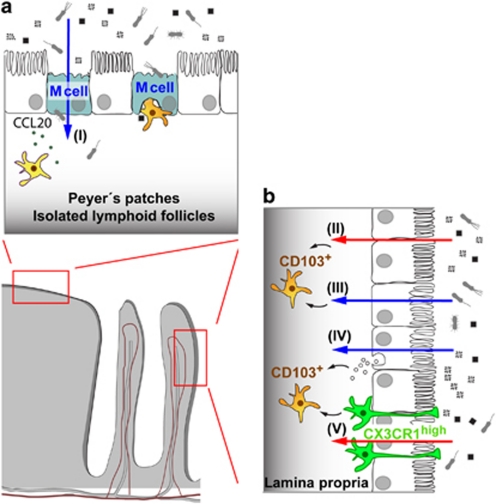
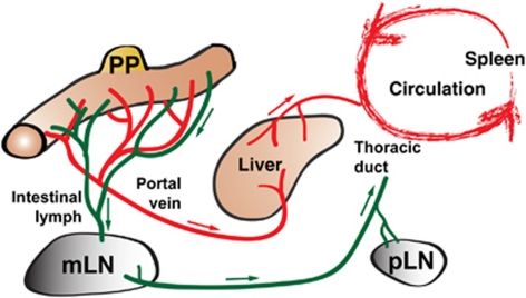
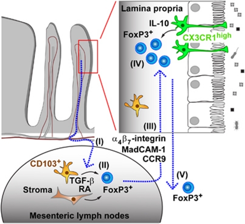
Oral tolerance is the state of local and systemic immune unresponsiveness that is induced by oral administration of innocuous antigen such as food proteins. An analogous but more local process also regulates responses to commensal bacteria in the large intestine and, together, mucosally induced tolerance appears to prevent intestinal disorders such as food allergy, celiac disease, and inflammatory bowel diseases. Here we discuss the anatomical basis of antigen uptake and recognition in oral tolerance and highlight possible mechanisms underlying the immunosuppression. We propose a model of stepwise induction of oral tolerance in which specialized populations of mucosal dendritic cells and the unique microenvironment of draining mesenteric lymph nodes combine to generate regulatory T cells that undergo subsequent expansion in the small intestinal lamina propria. The local and systemic effects of these regulatory T cells prevent potentially dangerous hypersensitivity reactions to harmless antigens derived from the intestine and hence are crucial players in immune homeostasis.


Figures

Figure 1
Figure 1
Mechanisms of antigen uptake in gut-associated lymphoid tissue (GALT) and lamina propria. (a) In organized tissues of the GALT such as Peyer's patches and isolated lymphoid follicles, specialized microfold cells (M cells) in the epithelium overlying the lymphoid follicles mediate transcellular transport of particulate material including intestinal microbiota (I). This antigen is then passed on to dendritic cells (DCs) that lie either below the epithelium or in a “pocket” created at the basolateral surface of the M cell. Production of CCL20 by surrounding epithelial cells can attract further DCs via their expression of CCR6. Some DCs in the GALT may also take up antigen directly from the lumen. (b) CD103+ DCs in the lamina propria underlying normal villus epithelium also play a critical role in presenting antigen for the induction of tolerance. They may acquire soluble antigens that have diffused through epithelial tight junctions (II), or that have been transferred across epithelial cells by transcellular routes (III). Exosomes containing antigen derived from class II MHC+ enterocytes may be taken up by DCs (IV). CX3CR1high macrophages have also been reported to capture luminal antigens by extending processes through the epithelial layer and they may pass this on to neighboring CD103+ DCs (V). There may also be a few M cells within villus epithelium (not depicted).
Figure 2
Figure 2
Routes by which intestinally derived antigen might disseminate and generate widespread systemic tolerance. Antigen taken up into Peyer's patches (PP) or lamina propria may enter the bloodstream via the portal vein, first reaching the liver before it becomes distributed into the wider circulation. Systemic tolerance may then occur because of antigen presentation in the liver by sinusoidal endothelial cells, tolerogenic conventional dendritic cells (DCs) or plasmacytoid DCs (pDCs), or because antigen reaching peripheral lymph nodes (pLNs) will be presented by resident DCs in the absence of costimulation. Free antigen taken up into afferent intestinal lymph will pass through the mesenteric lymph nodes (mLNs) and eventually enter the bloodstream via the thoracic duct.
Figure 3
Figure 3
A multistep model of oral tolerance to soluble antigens. Oral tolerance is initiated by the migration of antigen-loaded CD103+ dendritic cells (DCs) from the lamina propria (LP) into the draining mesenteric lymph nodes (mLNs; I). In the mLN, retinoic acid (RA) produced by DCs and local stromal cells induces the expression of gut-homing receptors α4β7 integrin and CCR9 on antigen-specific T cells and favors the transforming growth factor-β (TGF-β)-dependent differentiation of Foxp3+ induced regulatory T cells (iTregs; II). These committed Tregs home back to the intestinal lamina propria (III), where they undergo secondary expansion under the influence of interleukin-10 (IL-10) produced by CX3CR1high macrophages in the lamina propria (IV). In a putative fifth step (V), some of the Tregs may exit from the mucosa via lymph and/or bloodstream, disseminating throughout the immune system, and promoting the systemic consequences of oral tolerance. Foxp3, forkhead box P3; MadCAM-1, mucosal vascular addressin cell adhesion molecule 1.

References

    1. Molodecky N.A., et al. Increasing incidence and prevalence of the inflammatory bowel diseases with time, based on systematic review. Gastroenterology. 2011;142,:46–54.e42. - PubMed
    1. Meresse B., Ripoche J., Heyman M., Cerf-Bensussan N. Celiac disease: from oral tolerance to intestinal inflammation, autoimmunity and lymphomagenesis. Mucosal Immunol. 2009;2,:8–23. - PubMed
    1. Wells H.G., Osborne T.B. The biological reactions of the vegetable proteins. I: Anaphylaxis. J. Infect. Dis. 1911;8,:66–124.
    1. Weiner H.L., da Cunha A.P., Quintana F., Wu H. Oral tolerance. Immunol. Rev. 2011;241,:241–259. - PMC - PubMed
    1. Faria A.M., Weiner H.L. Oral tolerance: therapeutic implications for autoimmune diseases. Clin. Dev. Immunol. 2006;13,:143–157. - PMC - PubMed

Publication types

Substances