. 2012 Mar;139(5):1023-33.
doi: 10.1242/dev.071555.

Initiation of synapse formation by Wnt-induced MuSK endocytosis

Affiliations

Initiation of synapse formation by Wnt-induced MuSK endocytosis

Laura R Gordon et al. Development. 2012 Mar.

Abstract

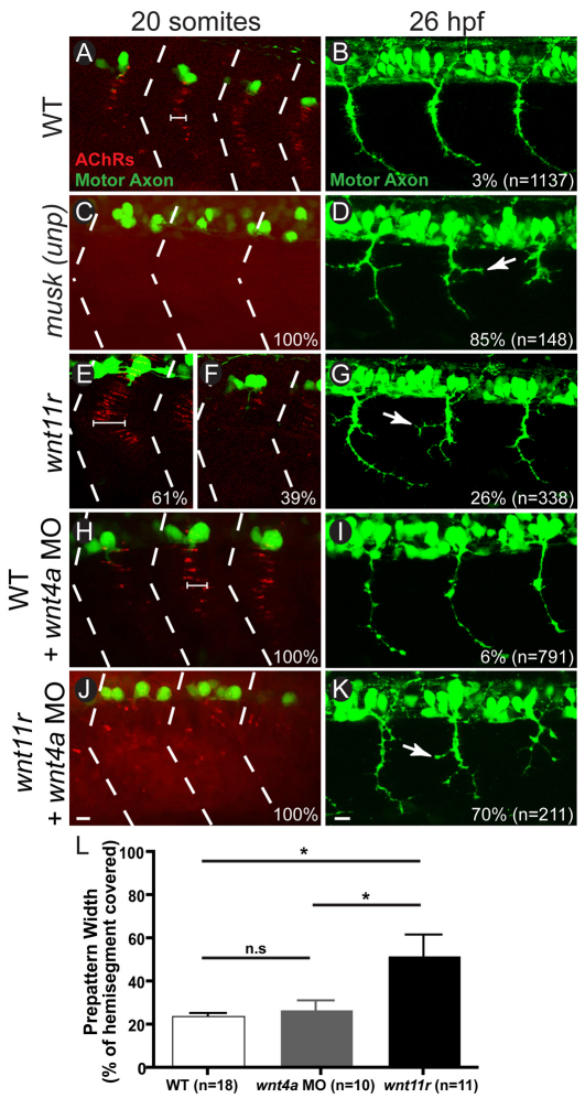
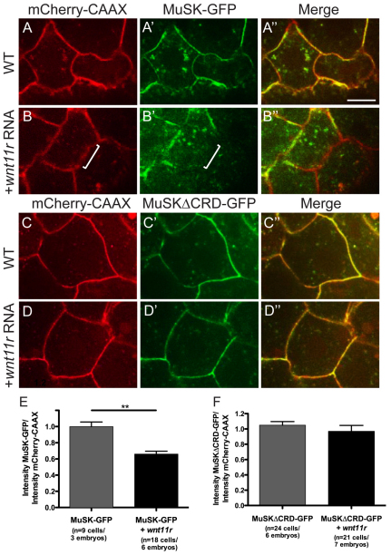
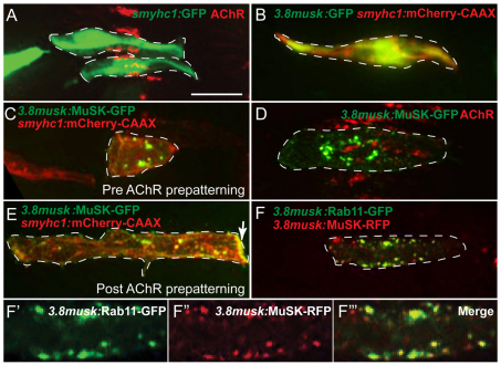
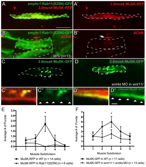
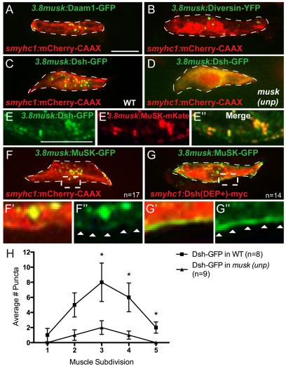
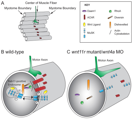
In zebrafish, the MuSK receptor initiates neuromuscular synapse formation by restricting presynaptic growth cones and postsynaptic acetylcholine receptors (AChRs) to the center of skeletal muscle cells. Increasing evidence suggests a role for Wnts in this process, yet how muscle cells respond to Wnt signals is unclear. Here, we show that in vivo, wnt11r and wnt4a initiate MuSK translocation from muscle membranes to recycling endosomes and that this transition is crucial for AChR accumulation at future synaptic sites. Moreover, we demonstrate that components of the planar cell polarity pathway colocalize to recycling endosomes and that this localization is MuSK dependent. Knockdown of several core components disrupts MuSK translocation to endosomes, AChR localization and axonal guidance. We propose that Wnt-induced trafficking of the MuSK receptor to endosomes initiates a signaling cascade to align pre- with postsynaptic elements. Collectively, these findings suggest a general mechanism by which Wnt signals shape synaptic connectivity through localized receptor endocytosis.


Figures

Fig. 1.
Fig. 1.
Loss of wnt11r and wnt4a phenocopies musk (unp) mutant. (A-K) Lateral view of trunk (top, dorsal; left, anterior) stained for AChR clusters (bungarotoxin, red) and motor axons (znp-1, green) in 20-somite and 26 hpf zebrafish embryos. (A,B) Three hemisegments in a wild-type embryo (hemisegment boundaries marked with dashed lines) with pre-patterned AChR clusters along the center of each hemisegment (cluster width marked by bracket; A) and three wild-type hemisegments in which motor axons have made synaptic contacts along the length of the trunk (B). (C,D) Three hemisegments in a musk (unp) mutant showing the absence of clustered AChRs, resulting in a complete dispersion of AChRs (C) and axon guidance errors in 26 hpf musk (unp) mutants (arrow points to axon branch) which occur in 85% of axons (D). (E-G) A single hemisegment from a wnt11r embryo showing an expanded AChR pre-pattern (width bracketed, 61% occurrence; E), and a single hemisegment showing a complete loss of AChR pre-pattern (39% of occurrence; F). Three hemisegments in a 26 hpf wnt11r mutant showing musk (unp)-like axon branching (arrow) in 26% of axons (G). (H,I) Three hemisegments in a wild-type embryo injected with 10 ng of wnt4a morpholino showing wild-type-like pre-patterning (H) and wild-type-like axons with only 6% guidance errors (I). (J,K) Three hemisegments from a wnt11r mutant injected with 10 ng of wnt4a morpholino showing musk (unp)-like AChR dispersal in all hemisegments (J) and guidance errors in 70% of axons (K). (L) Quantification of increase in AChR pre-pattern width in wnt11r mutants compared with wild type or wnt4a morpholino injected (P=0.01 for wild type vs wnt11r mutant, P=0.02 for wnt11r mutant vs wnt4a morpholino injected and P=0.38 for wild type vs wnt4a morpholino injected; Student’s two-tailed t-test, unequal variance; error bars indicate s.e.m.). Scale bars: 10 μm.
Fig. 2.
Fig. 2.
Addition of wnt11r triggers a loss of MuSK-GFP at the membrane in a CRD-dependent manner. (A-D″) Dome-stage zebrafish embryos at 4.5 hpf expressing mCherry-CAAX membrane marker (red) and either MuSK-GFP or MuSKΔCRD-GFP (green) in separate and merged channel views. (A-A″) Wild-type embryos showing MuSK-GFP membrane localization. (B-B″) Wild-type embryos injected with wnt11r mRNA showing reduced MuSK-GFP at the membrane (white bracket highlights one area of reduced GFP signal). (C-C″) Wild-type embryos showing MuSKΔCRD-GFP expression at the membrane, which is not reduced in the presence of wnt11r mRNA (D-D″). (E) Quantification of effect of wnt11r on MuSK-GFP membrane localization (**P=4.6×10⁻⁵; Student’s two-tailed t-test, unequal variance). (F) Quantification of effect of wnt11r on MuSKΔCRD-GFP membrane localization (P=0.39; Student’s two-tailed t-test, unequal variance). Error bars represent s.e.m. Scale bar: 10 μm.
Fig. 3.
Fig. 3.
MuSK-GFP localizes to Rab11-positive endosomes in the center of presynaptic muscle cells. (A) Expression of GFP driven by the smyhc1 promoter in two wild-type adaxial muscle cells (green) with pre-patterned AChR clusters (bungarotoxin, red) along the center of adaxial muscle cells. (B) Expression of 3.8musk:GFP (green) and membrane marker smyhc1:mCherry-CAAX (red) in an individual adaxial muscle cells. (C) 3.8musk:MuSK-GFP (green) and smyhc1:mCherry-CAAX (red) in a ‘young’ adaxial muscle cell that does not yet have pre-patterned AChR clusters. (D) 3.8musk:MuSK-GFP in an adaxial muscle cell (green) with AChRs (bungarotoxin, red) showing that MuSK-GFP puncta aggregate in close proximity to pre-patterned AChR clusters. (E) 3.8musk:MuSK-GFP (green) and smyhc1:mCherry-CAAX (red) in an ‘older’ adaxial muscle cell that no longer has pre-patterned AChRs, showing a reduction in central MuSK-GFP puncta (accumulation of protein at the myoseptal boundary is marked with an arrow). (F-F″′) A single confocal slice showing colocalization of MuSK-RFP (red) and early/recycling endosome marker Rab11-GFP (green) expressed under the 3.8musk promoter in a normal view (F) and magnified views (F-F″). Dashed lines encircle a single muscle cell. Scale bar: 10 μm.
Fig. 4.
Fig. 4.
MuSK trafficking is disrupted in the absence of rab11 and wnt11r/4a. (A,A′) Co-expression of smyhc1:rab11(S25N)-GFP (green) and 3.8musk:MuSK-RFP (red; alone in A′), demonstrating disruption of MuSK-RFP center muscle localization. Instead, MuSK-RFP appears dispersed throughout the cell. (B,B′) smyhc1:rab11(S25N)-GFP (green) expressed in two adjacent muscle cells showing disruption of AChR clusters (bungarotoxin, red) at the interface of the two mosaic fibers but not in an adjacent wild-type cell (arrow). Observed in 38% of cell interfaces, n=13. (C-C″) 3.8musk:MuSK-GFP (green) in a wild-type embryo with normal protein distribution (C) and a magnified view of dashed box showing very little protein at the membrane as visualized with smyhc:mCherryCAAX (C′,C″). (D-D″) 3.8musk:MuSK-GFP (green) in a wnt11r mutant/wnt4a morphant embryo (D), and magnified view of dashed box showing accumulation of protein at the membrane as visualized with smyhc:mCherryCAAX (D′,D″). (E,F) Quantification of central enrichment of MuSK-RFP puncta when co-expressed with Rab11(S25N)-GFP (E) (P values for muscle subdivisions 1-5: 0.6, 0.27, 0.01, 0.38, 0.34, respectively), or when expressed in a wnt11r mutant/wnt4a morphant background (F; P values for muscle subdivisions 1-5: 0.87, 0.12, 0.04, 0.36, 0.97, respectively). *P<0.05. Error bars represent s.e.m. Dashed lines encircle a single muscle cell. Arrowheads (C″,D″) indicate cell membrane. Scale bars: 10 μm in A-D; 1 μm in C′-D″.
Fig. 5.
Fig. 5.
Localization of noncanonical PCP proteins to the center of muscle cells requires MuSK, and vice versa. (A,B) Daam1-GFP (green) (A) and Diversin-YFP (green) (B) under the 3.8musk promoter localize to centrally enriched puncta in muscle cells co-expressing mCherry-CAAX under the smyhc1 promoter (red). (C) Dsh-GFP under the 3.8musk promoter (green) localizes to centrally enriched puncta in fibers co-expressing mCherry-CAAX (red). (D) The punctate localization of Dsh-GFP (green) is lost in musk (unp) mutant muscle cells (mCherry-CAAX in red). (E-E″) Magnified view of center of muscle cell showing colocalization of Dsh-GFP (green) and MuSK-mKate (red), both driven by the 3.8musk promoter. (F-F″) MuSK-GFP under the 3.8musk promoter (green) localizes to centrally enriched puncta in muscle cells co-expressing mCherry-CAAX (red). F′ and F″ show magnified views of the region marked with dashed box in F. (G-G″) MuSK-GFP localizes to the membrane of muscle cells co-expressing Dsh(DEP+)-Myc. G′ and G″ show magnified views of the region marked with dashed box in G. (H) Quantification of reduction in centrally localized Dsh-GFP puncta in musk (unp) mutants (P values for muscle subdivisions 1-5: 0.49, 0.09, 0.05, 0.04, 0.02, respectively). *P<0.05. Error bars represent s.e.m. Dashed lines encircle a single muscle cell. Arrowheads in F″ and G″ indicate cell membrane. Scale bars: 10 μm.
Fig. 6.
Fig. 6.
Model for Wnt-dependent receptor endocytosis directing synapse position. (A) Schematic of a somitic hemisegment with parallel early muscle cells in gray. AChRs (red) are clustered in the center of the muscle fibers along the path where the motor axon (green) will extend. (B) Magnified view of dashed box in A showing a cross-section of a single wild-type muscle fiber. Motor axon growth cones (green) are navigating in the center of cells, precisely where AChRs (red) are clustered. In a Wnt ligand (yellow)-dependent manner, MuSK (blue) translocates from the membrane into Rab11-positive endosomes restricted to the center of muscle cells. It is unclear whether Wnt11r and Wnt4a directly activate the MuSK receptor in vivo or act indirectly on MuSK trafficking. Once internalized, the cytoplasmic domain of MuSK binds Dishevelled (orange) and nucleates accumulation of Diversin (Ankrd6; brown), Daam1 (purple) and RhoA (green) to modify the cytoskeleton, ultimately anchoring AChRs to the cell center. (C) Magnified view of showing a cross-section of a wnt11r mutant/wnt4a morphant muscle cell, in which MuSK, Dishevelled, Daam1, Diversin and RhoA are no longer localized in the center. Consequently, AChRs and growth cones (green) are no longer restricted to the muscle center.

References

    1. Alberts A. S. (2002). Diaphanous-related Formin homology proteins. Curr. Biol. 12, R796 - PubMed
    1. Axelrod J. D., Miller J. R., Shulman J. M., Moon R. T., Perrimon N. (1998). Differential recruitment of Dishevelled provides signaling specificity in the planar cell polarity and Wingless signaling pathways. Genes Dev. 12, 2610–2622 - PMC - PubMed
    1. Banerjee S., Gordon L., Donn T. M., Berti C., Moens C. B., Burden S. J., Granato M. (2011). A novel role for MuSK and non-canonical Wnt signaling during segmental neural crest cell migration. Development 138, 3287–3296 - PMC - PubMed
    1. Bruneau E. G., Akaaboune M. (2006). The dynamics of recycled acetylcholine receptors at the neuromuscular junction in vivo. Development 133, 4485–4493 - PubMed
    1. Budnik V., Salinas P. C. (2011). Wnt signaling during synaptic development and plasticity. Curr. Opin. Neurobiol. 21, 151–159 - PMC - PubMed

Publication types

MeSH terms