. 2012 Apr 27;287(18):14659-71.
doi: 10.1074/jbc.M111.316323. Epub 2012 Feb 8.

Rpn1 and Rpn2 coordinate ubiquitin processing factors at proteasome

Affiliations

Rpn1 and Rpn2 coordinate ubiquitin processing factors at proteasome

Rina Rosenzweig et al. J Biol Chem. .

Abstract

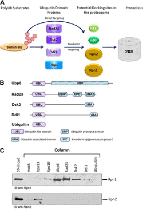
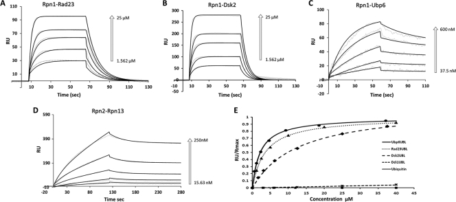
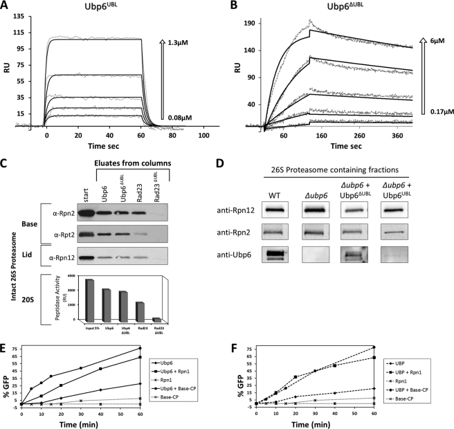
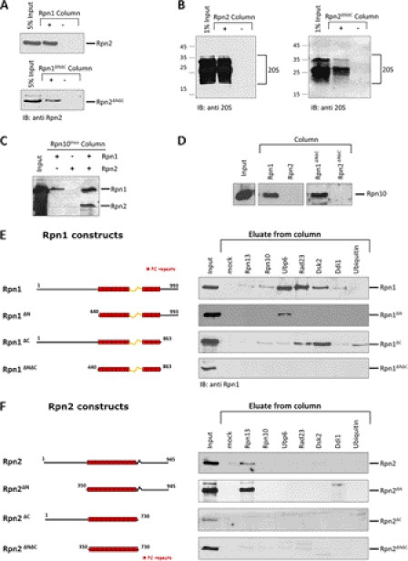
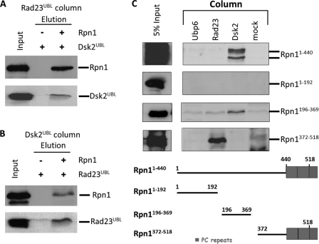
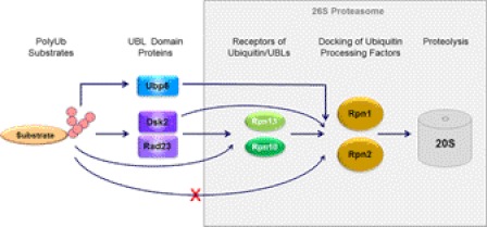
Substrates tagged with (poly)ubiquitin for degradation can be targeted directly to the 26 S proteasome where they are proteolyzed. Independently, ubiquitin conjugates may also be delivered by bivalent shuttles. The majority of shuttles attach to the proteasome through a ubiquitin-like domain (UBL) while anchoring cargo at a C-terminal polyubiquitin-binding domain(s). We found that two shuttles of this class, Rad23 and Dsk2, dock at two different receptor sites embedded within a single subunit of the 19 S proteasome regulatory particle, Rpn1. Their association/dissociation constants and affinities for Rpn1 are similar. In contrast, another UBL-containing protein, the deubiquitinase Ubp6, is also anchored by Rpn1, yet it dissociates slower, thus behaving as an occasional proteasome subunit that is distinct from the transiently associated shuttles. Two neighboring subunits, Rpn10 and Rpn13, show a marked preference for polyubiquitin over UBLs. Rpn10 attaches to the central solenoid portion of Rpn1, although this association is stabilized by the presence of a third subunit, Rpn2. Rpn13 binds directly to Rpn2. These intrinsic polyubiquitin receptors may compete with substrate shuttles for their polyubiquitin-conjugate cargos, thereby aiding release of the emptied shuttles. By binding multiple ubiquitin-processing factors simultaneously, Rpn1 is uniquely suited to coordinate substrate recruitment, deubiquitination, and movement toward the catalytic core. The broad range of affinities for ubiquitin, ubiquitin-like, and non-ubiquitin signals by adjacent yet nonoverlapping sites all within the base represents a hub of activity that coordinates the intricate relay of substrates within the proteasome, and consequently it influences substrate residency time and commitment to degradation.


Figures

FIGURE 1.
FIGURE 1.
Rpn1 and Rpn2 dock ubiquitin processing factors. A, a number of UDPs serve as bifunctional shuttles by interacting simultaneously with polyubiquitinated cargo and with receptors at the proteasome. PolyUb conjugates can also dock at the proteasome directly. Four proteasome subunits (Rpn1, Rpn2, Rpn10, and Rpn13) have been implicated in recruiting ubiquitin chains or UBLs. B, most UDPs contain an N-terminal UBL followed by a unique functional domain. Some bind polyUb through a UBA. Rad23 contains two UBA domains, UBA1 and UBA2, which share 27% sequence identity, and an XPC (xeroderma pigmentosum group C complementing protein) domain between the two (92). Dsk2 and Ddi1 contain only one UBA domain, with Ddi1 also having an internal retroviral aspartyl protease domain (93). Ubp6 associates with ubiquitin via its UBP. Protein domains are drawn roughly to scale. C, immobilized ubiquitin processing factors (Rpn13, Rpn10, Ubp6, Rad23, Dsk2, and Ddi1) as well as ubiquitin were each tested for binding Rpn1 or Rpn2 proteins. Rpn is regulatory particle non-ATPase. IB, immunoblot.
FIGURE 2.
FIGURE 2.
Binding kinetics to Rpn1 or Rpn2. Rpn1 and Rpn2 were immobilized on separate channels (1700 and 1100 response units (RU)) of a ProteOn surface plasmon resonance sensor and injected with increasing concentrations of their potential binding partners followed by buffer wash. Positive associations are displayed for Rpn1-Rad23 (A), Rpn1-Dsk2 (B), Rpn1-Ubp6 (C), and Rpn2-Rpn13 (D). The response data (dashed lines) for association and dissociation are shown for a series of concentrations (arrows) overlaid with a model fit (solid line). Sensograms in A, B, and D were fit to the Langmuir model for 1:1 binding stoichiometry. Sensograms in C (Rpn1-Ubp6 response) were fit to an HLPR, assuming two-binding sites. Association (kon) and dissociation (koff) rate constants derived from a global fit of primary response data over the entire concentration range for each pair to the corresponding model are summarized in Table 1. E, binding isotherms for Ubp6UBL (circles), Rad23UBL (triangles), Dsk2UBL (diamonds), Ddi1UBL (×), and ubiquitin (squares) derived from equilibrium SPR measurements. Normalized equilibrium response is plotted as a function of soluble protein concentration; the fitting curves correspond to dissociation constants shown in Table 2.
FIGURE 3.
FIGURE 3.
Two-step proteasome incorporation of Ubp6. A and B, SPR sensograms of Ubp6UBL and Ubp6ΔUBL binding kinetics to Rpn1. The response data for binding and dissociation (dotted lines) are shown for a series of concentrations (arrows). Values of best fit parameters to a single bimolecular interaction model (solid lines) are summarized in Table 3. RU, response units. C, immobilized Ubp6 and Rad23 lacking their UBL domains (Ubp6ΔUBL and Rad23ΔUBL) were tested for binding to 26 S proteasome-purified free of all UDPs or other transient proteasome-interacting proteins. Association of full-length Ubp6 and Rad23 is included as positive control. Eluate from each column was tested for proteasome activity (20 S CP) as well as immunoblotted (IB) for presence of 19 S subunits from the Base and Lid subcomplexes. D, truncated Ubp6-expressing strains were molecular weight-fractionated, and proteins copurifying with peak of proteasome activity were identified by immunoblotting. E, potency of Ubp6 to remove ubiquitin from Ub-GFP was monitored over 60 min, and % reaction progression was plotted over time (diamonds). The reaction was repeated in presence of equimolar Rpn1 (squares) or purified base-CP (lidless) proteasome lacking both endogenous Ubp6 and Rpn11 (circles). Neither Rpn1 (star) nor base-CP alone (×) exhibit deubiquitination activity in the absence of added Ubp6 (dotted lines). F, experiment similar to E but for truncated Ubp6ΔUBL. Ubp6 activation is not dependent on UBL.
FIGURE 4.
FIGURE 4.
Mapping binding sites on Rpn1 and Rpn2. Indicated proteins were immobilized on CH-Sepharose beads, mixed with various fragments of Rpn1 or Rpn2 proteins, washed, and eluates analyzed by immunoblotting (IB) for retained Rpn1 or Rpn2.
FIGURE 5.
FIGURE 5.
Dsk2 and Rad23 bind to two different sites at Rpn1. A and B, immobilized Rad23UBL was incubated with either Dsk2UBL or a premixed Rpn1 + Dsk2UBL mixture and washed, and bound proteins were eluted and immunoblotted for the presence of Dsk2. The reciprocal experiment was repeated with Dsk2UBL immobilized and incubated with either Rad23UBL or a Rpn1 + Rad23UBL mixture. The formation of these Rad23-Rpn1-Dsk2 ternary complexes demonstrates that Rad23 and Dsk2 can bind simultaneously to Rpn1, via at least two discrete binding sites. C, purified recombinant N-terminal fragments of Rpn1 were washed over immobilized binding partners, washed, and elution immunoblotted for presence of Rpn1. A Dsk2-binding site is located between residues 196 and 369 of Rpn1, whereas Rad23 binds tighter to an Rpn1 fragment spanning residues 372–518.
FIGURE 6.
FIGURE 6.
Schematic description of substrate relay. Polyubiquitin-binding proteins such as Dsk2 or Rad23 are transiently associated with proteasomes and thus may serve as shuttles or delivery proteins for polyubiquitin conjugates. These proteins contain an N-terminal UBL (ubiquitin-like) domain through which they dock at the proteasome. The primary UBL receptors for Rad23 and Dsk2 was found to be proteasome subunits Rpn1 (Tables 1–3), although these two UDPs (UBL-domain containing proteins) were also capable of associating directly with two neighboring subunits, Rpn10 and Rpn13 (supplemental Table ST1). Ddi1 apparently also associates with Rpn1, although in this study we were unable to quantify the intensity of their mutual association. Polyubiquitin chains could also bind to Rpn10 and Rpn13 directly (supplemental Table ST1), thereby bypassing the need for shuttles. The relative affinities of these receptors for the different (poly)ubiquitin or UBL signals influence efficiencies of substrate targeting and coordinate their entry into the proteasome. Another UDP, the deubiquitinating enzyme Ubp6, bound only to Rpn1 yet much tighter than transiently associated shuttle proteins, thus behaving more akin to proteasome subunits Rpn10 and Rpn13. The proximal localization of Ubp6 to these polyubiquitin-chain-receptors may also coordinate trimming, processing, or shaving of conjugated chains with substrate preparation and the proteolytic mechanism (see additional details in supplemental Fig. S1).

References

    1. Finley D. (2009) Recognition and processing of ubiquitin-protein conjugates by the proteasome. Annu. Rev. Biochem. 78, 477–513 - PMC - PubMed
    1. Bedford L., Paine S., Sheppard P. W., Mayer R. J., Roelofs J. (2010) Assembly, structure, and function of the 26 S proteasome. Trends Cell Biol. 20, 391–401 - PMC - PubMed
    1. Glickman M. H., Rubin D. M., Coux O., Wefes I., Pfeifer G., Cjeka Z., Baumeister W., Fried V. A., Finley D. (1998) A subcomplex of the proteasome regulatory particle required for ubiquitin-conjugate degradation and related to the COP9-signalosome and eIF3. Cell 94, 615–623 - PubMed
    1. Guterman A., Glickman M. H. (2004) Complementary roles for Rpn11 and Ubp6 in deubiquitination and proteolysis by the proteasome. J. Biol. Chem. 279, 1729–1738 - PubMed
    1. Bar-Nun S., Glickman M. H. (2011) Proteosomal AAA-ATPases. Structure and function. Biochim. Biophys. Acta 1823, 67–82 - PubMed

Publication types

MeSH terms

LinkOut - more resources