. 2012 May;26(5):2137-44.
doi: 10.1096/fj.11-201640. Epub 2012 Feb 7.

Anti-inflammatory effects of β2 adrenergic receptor agonists in experimental acute lung injury

Affiliations

Anti-inflammatory effects of β2 adrenergic receptor agonists in experimental acute lung injury

Markus Bosmann et al. FASEB J. 2012 May.

Abstract

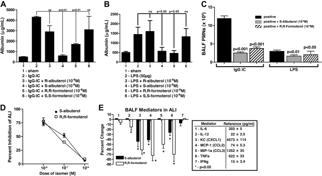
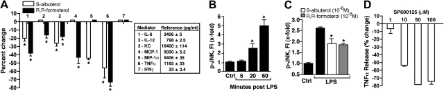
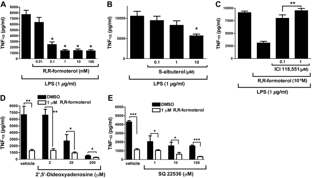
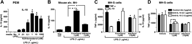
These studies were undertaken to extend emerging evidence that β(2) adrenergic receptor (β(2)AR) agonists, in addition to their bronchorelaxing effects, may have broad anti-inflammatory effects in the lung following onset of experimental acute lung injury (ALI). Young male C57BL/6 mice (25 g) developed ALI following airway deposition of bacterial LPS or IgG immune complexes in the absence or presence of appropriate stereoisomers (enantiomers) of β(2)AR agonists, albuterol or formoterol. Endpoints included albumin leak into lung and buildup of polymorphonuclear neutrophils and cytokines/chemokines in bronchoalveolar fluids. Both β(2)AR agonists suppressed lung inflammatory parameters (IC(50)=10(-7) M). Similar effects of β(2)AR agonists on mediator release were found when mouse macrophages were stimulated in vitro with LPS. The protective effects were associated with reduced activation (phosphorylation) of JNK but not of other signaling proteins. Collectively, these data suggest that β(2)AR agonists have broad anti-inflammatory effects in the setting of ALI. While β(2)AR agonists suppress JNK activation, the extent to which this can explain the blunted lung inflammatory responses in the ALI models remains to be determined.


Figures

Figure 1.
Figure 1.
Effects of β2AR agonists during ALI. A) Acute lung injury in young adult (25 g) C57BL/6 male mice 6 h after intratracheal (i.t.) administration of 125 μg anti-BSA IgG and intravenous (i.v.) infusion of 1 mg BSA, resulting in IgGIC-induced ALI. Sham-treated mice received i.t. anti-BSA but no i.v. BSA. Isomeric forms (enantiomers) of β2AR agonists were intermixed with the anti-BSA, all at 10−6 M. For each bar, n ≥ 12 mice. Endpoints were BALF mouse albumin as measured by ELISA. B) ALI induced by i.t. administration of 50 μg LPS 6 h earlier, resulting in ALI. Various enantiomers of albuterol were used (as indicated), at 10−6 M, similar to the protocol in panel A. For each bar, n ≥ 10 mice. Endpoints were mouse albumin in BALFs. C) Effects of 10−6 M S-albuterol or R,R-formoterol on BALF content of PMNs 6 h after onset of IgGIC-induced or LPS-induced ALI. Virtually no PMNs were found in sham-treated BALFs. For each bar, n ≥ 10. D) Dose responses of S-albuterol or R,R-formoterol (i.t.) on BALF albumin 6 h after onset of IgGIC-ALI. For each dose, n ≥ 5 mice. BALF albumin levels were compared as a percentage of albumin in positive controls. E) BALF mediators in IgGIC-ALI mice 6 h after onset of reactions, expressed as a percentage of positive controls (pg/ml; shown at right). S-albuterol or R,R-formoterol were present at 10−6 M. Positive control reference values (pg/ml) are indicated for each of the 7 mediators. For each bar, n ≥ 6 mice. *P < 0.05.
Figure 2.
Figure 2.
Effects of β2AR agonists and JNK inhibitors on mediator production in vitro. A) Effects of β2AR agonists on mediator production in LPS-stimulated (1 μg/ml) mouse PEMs (2×10⁶) for 4 h at 37°C, expressed as percentage reduction. Positive control reference values (pg/ml) are indicated for each of the 7 mediators. As indicated, 10−6 M S-albuterol or R,R-formoterol was present at the time of addition of LPS to PEMs. Percentage reduction is relative to LPS-stimulated PEM in the absence of β2AR agonists. Reference values (pg/ml) for positive controls are shown in box. For each bar, n = 6 samples. B) Activation (phosphorylation) of JNK in LPS-stimulated PEMs as a function of time, using control (ctrl) PEMs (non-LPS exposed) as the reference point. Phospho-JNK was measured in cell lysates as a function of time (min) after addition of LPS, using a bead-based assay. For each bar, n = 4 samples. C) Levels of phospho-JNK in LPS-stimulated PEMs in the absence or presence of 10−6 M S-albuterol or R,R-formoterol 60 min after addition of LPS ± β2AR agonists. Results are from 4 separate experiments, in which average reductions of JNK phosphorylation in the presence of S-albuterol or R,R-formoterol were 31 and 35%, respectively. For each bar, n ≥ 4 samples. D) Dose response for SP600125 (JNK1/2 inhibitor) on release of TNF-α from LPS-stimulated PEMs. For each bar, n ≥ 4 samples. *P < 0.05.
Figure 3.
Figure 3.
In vitro dose responses of LPS-stimulated PEMs in the absence or presence of β2AR agonists, using release of TNF-α as the endpoint. A) TNF-α release in the presence of increasing concentrations of R,R-formoterol (0.01–100 nM). B) Similar experiments using S-albuterol (0.1–10 μM). Text describes parallel effects on MCP-1, MIP-1α, KC, and IL-6. C) Similar experiments with R,R-formoterol (1 μM) in the absence or presence of ICI-118,551 (selective β2AR antagonist). D) Release of TNF-α from PEMs activated with LPS (1 μg/ml) for 4 h at 37°C in the absence or presence of 1 μM R,R-formoterol and, as indicated, in the copresence of 1–200 μM 2′,5′dideoxyadenosine (inhibitor of adenylate cyclase). E) Experiments similar to those in D, using another adenylate cyclase inhibitor (SQ2536) at concentrations of 1–100 μM. For all bars, n ≥ 5 samples. *P < 0.05, **P < 0.01, ***P < 0.001.
Figure 4.
Figure 4.
Suppression of TNF-α production by β2AR agonists is independent of IL-10. A, B) Production of IL-10 by LPS-stimulated mouse PEMs (A) and mouse alveolar macrophages (MΦ; B). Cells were stimulated by LPS (1 μg/ml) for 4 h in the absence or presence of R,R-formoterol or S-albuterol (both at 10−6 M). C) Production of TNF-α (solid bars, left y axis) and IL-10 (open bars, right y axis) in LPS-stimulated MH-S cells (immortalized mouse lung macrophages) in the presence or absence of 1 × 10⁻⁶ M R,R-formoterol or S-albuterol, 4 h. D) Similarly treated cells were also exposed to LPS in the presence or absence of 2 different concentrations of neutralizing antibody to IL-10 at the indicated concentrations. For all bars, n ≥ 5 samples. *P < 0.05.
Figure 5.
Figure 5.
Histopathology of mouse lungs in normal lungs and lungs after ALI following airway deposition of IgGICs. A) Histological features of normal mouse lung. B) Positive control (acutely injured lung induced by airway deposition of IgGICs) 6 h after induction of ALI. C, D) Lungs after initiation of ALI in the copresence of 10−6 M S-albuterol (C) or R,R-formoterol (D). All panels are from paraffin-embedded sections stained with hematoxylin and eosin (×40 view). Insets: ×100 view.

References

    1. Matthay M. A., Zimmerman G. A. (2005) Acute lung injury and the acute respiratory distress syndrome: four decades of inquiry into pathogenesis and rational management. Am. J. Respir. Cell Mol. Biol. 33, 319–327 - PMC - PubMed
    1. Goss C. H., Brower R. G., Hudson L. D., Rubenfeld G. D. (2003) Incidence of acute lung injury in the United States. Crit. Care Med. 31, 1607–1611 - PubMed
    1. Sakuma T., Folkesson H. G., Suzuki S., Okaniwa G., Fujimura S., Matthay M. A. (1997) Beta-adrenergic agonist stimulated alveolar fluid clearance in ex vivo human and rat lungs. Am. J. Respir. Crit. Care Med. 155, 506–512 - PubMed
    1. Sakuma T., Okaniwa G., Nakada T., Nishimura T., Fujimura S., Matthay M. A. (1994) Alveolar fluid clearance in the resected human lung. Am. J. Respir. Crit. Care Med. 150, 305–310 - PubMed
    1. Tracey K. J. (2009) Reflex control of immunity. Nat. Rev. Immunol. 9, 418–428 - PMC - PubMed

Publication types

Substances