Review
. 2010;10(4):3218-42.
doi: 10.3390/s100403218. Epub 2010 Apr 1.

Visual pathways serving motion detection in the mammalian brain

Affiliations
Review

Visual pathways serving motion detection in the mammalian brain

Alice Rokszin et al. Sensors (Basel). 2010.

Abstract

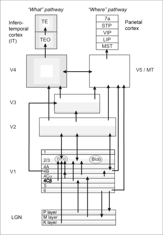
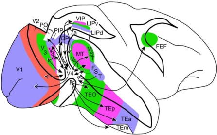
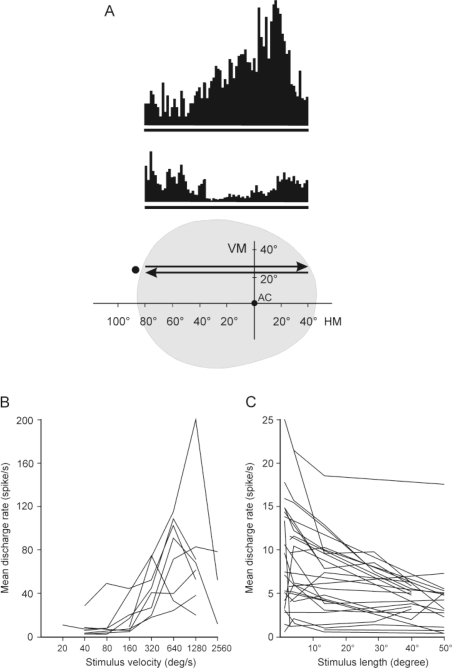
Motion perception is the process through which one gathers information on the dynamic visual world, in terms of the speed and movement direction of its elements. Motion sensation takes place from the retinal light sensitive elements, through the visual thalamus, the primary and higher visual cortices. In the present review we aim to focus on the extrageniculo-extrastriate cortical and subcortical visual structures of the feline and macaque brain and discuss their functional role in visual motion perception. Special attention is paid to the ascending tectofugal system that may serve for detection of the visual environment during self-motion.

Keywords: ascending tectofugal system; caudate nucleus; dorsal stream; motion detection; posterior thalamus; ventral stream.


Figures

Figure 1.
Figure 1.
Schematic representation of the primate visual pathways. The left half of the figure represents the ventral (“what”) stream, while the right side shows the hierarchical organization of the dorsal (“where”) stream. Abbreviations: LGN—Lateral geniculate nucleus, V1, V2, V3, V4—Primary (first), second, third, fourth visual cortices, respectively, TEO—Posterior inferior temporal cortex, TE—Anterior inferior temporal cortex, V5/MT—Middle temporal area (fifth visual cortex), MST—Medial superior temporal area, LIP—Lateral intraparietal area, VIP—Ventral intraparietal area, STP—Superior temporal polysensory area, 7a—Visual area 7a in the parietal cortex (Brodmann’s terminology).
Figure 2.
Figure 2.
Location of the cortical visual areas in the primate. Open arrowheads indicate feedback projections, closed arrowheads indicate feedforward projections. Occipital areas: V1, V2, V3, V4—Primary (first), second, third, fourth visual cortices respectively, Temporal areas: TEO—Posterior inferior temporal cortex, TE—Anterior inferior temporal cortex, MT—Middle temporal area (fifth visual cortex), MST—Medial superior temporal area, FST—Fundus of the superior temporal area, Parietal areas: LIP—Lateral intraparietal area, VIP—Ventral intraparietal area, PIP—Posterior intraparietal area, PO—parieto-occipital sulcus, 7a—Visual area 7a in the parietal cortex (Brodmann’s terminology), Frontal area: FEF—Frontal eye field. According to the figure of Ungerleider et al. [73].
Figure 3.
Figure 3.
Visual pathways in the feline brain. This schematic figure shows the geniculo-cortical (primary) visual pathway (blue arrows) and the ascending tectofugal visual pathway (red arrows) in the cat’s brain. Abbreviations: LGN—Lateral geniculate nucleus, Pul—Pulvinar, LP—Lateral-posterior nucleus of the thalamus, A17—Visual area 17 (primary visual cortex), LS—Lateral suprasylvian cortex, AEV—anterior ectosylvian visual area, IVA—Insular visual area, LM-Sg—Lateral medial-suprageniculate nuclei of the thalamus, SNr—Substantia nigra pars reticulata, PPT—Pedunculo-pontin tegmental nuclei, STN—Subthalamic nucleus, SCs, SCi, SCd—Superior colliculus (superficial, intermedier, deep layers, respectively), CN—Caudate nucleus, FEF—Frontal eye field.
Figure 4.
Figure 4.
Visual properties of neurons in the ascending tectofugal system. A: Top: Peristimulus time histograms (PSTHs) of a directionally selective LM-Sg visual neuron in the ascending tectofugal system. Bottom: the position and movement of the stimulus in the visual field of the cat. The black spot left to the arrows symbolizes the moving visual stimulus. The upper and lower PSTHs correspond to the response of the neuron to the stimulus moving along the trace indicated by the upper and lower arrows, respectively. The grey part represents the extent of the visual receptive field. Abbreviations: AC: area centralis, HM: horizontal meridian, VM: vertical meridian. B: Velocity response curves for a spot stimulus (2° in diameter) of eight visual AEV neurons of this system. Note the high responsiveness of the units to high velocities! C: Effect of the length of the light stimulus (1° in width) on the response of visual neurons in the AEV. Note the maximal responsiveness of the neurons to small stimuli!

References

    1. Nakayama K. Biological image motion processing: A review. Vis. Res. 1985;25:625–660. - PubMed
    1. Braunstein M.L. Sensitivity of the observer to transformations of the visual field. J. Exp. Psychol. 1966;72:638–687. - PubMed
    1. Simpson J.I., Leonard C.S., Soodak R.E. The accessory optic-system. Analyzer of self-motion. Ann. N. Y. Acad. Sci. 1988;545:170–179. - PubMed
    1. Goodale M.A., Milner A.D. Separate visual pathways for perception and action. Trends Neurosci. 1992;15:20–25. - PubMed
    1. Ungerleider L.G., Mishkin M. Two cortical visual systems. In: Ingle D.J., Goodale M.A., Mansfield R.J.W., editors. Analysis of Visual Behavior. MIT Press; Cambridge, MA, USA: 1982. pp. 549–586.

Publication types

LinkOut - more resources