Primary leiomyosarcoma of the pancreas
- PMID: 22319744
- PMCID: PMC3267071
- DOI: 10.4174/jkss.2011.81.Suppl1.S69
Primary leiomyosarcoma of the pancreas
Abstract
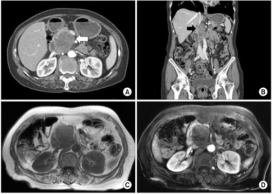
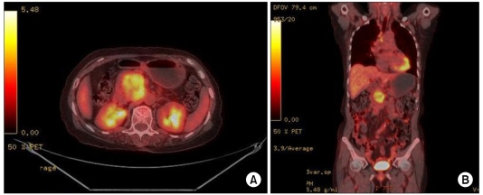
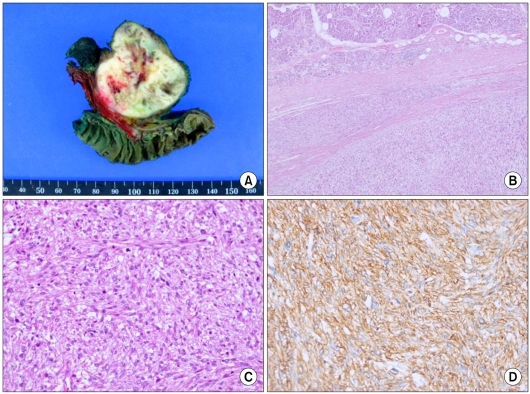
Primary sarcomas of the pancreas are extremely rare, accounting for 0.1% of malignant pancreatic (non-islet) neoplasms. Pancreatic leiomyosarcoma is a highly aggressive malignancy that spreads in a similar manner to gastric leiomyosarcoma, i.e., by adjacent organ invasion, hematogenous spread, and lymph node metastasis. These tumors are large at the time of diagnosis and are usually found at an advanced stage. We report a case of a 70-year-old female with intermittent right upper quadrant abdominal discomfort. Radiological, histopathological, and immunohistochemical studies revealed the tumor to be a primary leiomyosarcoma of the pancreas. Herein, we describe a patient with a primary leiomyosarcoma of the pancreas who presented with clinical and radiological findings indicative of a mass in the pancreatic head.
Keywords: Leiomyosarcoma; Pancreas; Primary.
Conflict of interest statement
No potential conflict of interest relevant to this article was reported.
Figures
References
-
- Baylor SM, Berg JW. Cross-classification and survival characteristics of 5,000 cases of cancer of the pancreas. J Surg Oncol. 1973;5:335–358. - PubMed
-
- Ross CF. Leiomyosarcoma of the pancreas. Br J Surg. 1951;39:53–56. - PubMed
-
- Komoda H, Nishida T, Yumiba T, Nishikawa K, Kitagawa T, Hirota S, et al. Primary leiomyosarcoma of the pancreas--a case report and case review. Virchows Arch. 2002;440:334–337. - PubMed
Publication types
LinkOut - more resources
Full Text Sources