. 2012 Mar;83(5):1035-47.
doi: 10.1111/j.1365-2958.2012.07987.x. Epub 2012 Feb 10.

The ancillary protein 1 of Streptococcus pyogenes FCT-1 pili mediates cell adhesion and biofilm formation through heterophilic as well as homophilic interactions

Affiliations
Free PMC article

The ancillary protein 1 of Streptococcus pyogenes FCT-1 pili mediates cell adhesion and biofilm formation through heterophilic as well as homophilic interactions

Marco Becherelli et al. Mol Microbiol. 2012 Mar.
Free PMC article

Abstract

Gram-positive pili are known to play a role in bacterial adhesion to epithelial cells and in the formation of biofilm microbial communities. In the present study we undertook the functional characterization of the pilus ancillary protein 1 (AP1_M6) from Streptococcus pyogenes isolates expressing the FCT-1 pilus variant, known to be strong biofilm formers. Cell binding and biofilm formation assays using S. pyogenes in-frame deletion mutants, Lactococcus expressing heterologous FCT-1 pili and purified recombinant AP1_M6, indicated that this pilin is a strong cell adhesin that is also involved in bacterial biofilm formation. Moreover, we show that AP1_M6 establishes homophilic interactions that mediate inter-bacterial contact, possibly promoting bacterial colonization of target epithelial cells in the form of three-dimensional microcolonies. Finally, AP1_M6 knockout mutants were less virulent in mice, indicating that this protein is also implicated in GAS systemic infection.


Figures

Fig 1
Fig 1
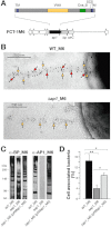
Structural organization and adhesive properties of the ancillary protein AP1_M6 of FCT-1 pili. A. Schematic representation of the FCT-1 pilus island genetic organization and of the structural domains of AP1_M6, including the hydrophobic transmembranes (TM), the Von-Willebrand factor type A domain (VWA), the Cna_B domain and the sortase cleavage site (SCS). B. Immunoelectron microscopy image taken at 30× magnification after double immunogold labelling of FCT-1 M6 pili from wild type (WT_M6) and its isogenic Δap1_M6 derivative with rabbit polyclonal anti-rBP_M6 and mouse anti-rAP1_M6 antibodies, followed by gold-labelled anti-rabbit IgG (particle size 5 nm, yellow arrows) and anti-mouse IgG antibodies (10 nm, red arrows). C. Immunoblot of cell wall fractions of WT_M6, Δap1_M6 and the complemented strain Δap1_M6(pAMap1_M6). Total protein amounts of the different extracts loaded on the gels were normalized using antibodies to protein Spy0269. Transferred extracts were incubated with specific mouse sera raised against rBP_M6 and rAP1_M6 proteins. D. Binding of WT_M6, Δap1_M6 and Δap1_M6(pAMap1_M6) to A549 cells (moi 100:1) after 30 min of incubation. Histograms represent the percentage of cell-associated versus total bacteria. Results are presented as the mean and standard deviation values of three independent experiments run in triplicate wells. Significant differences between strains calculated by Student's t-test are shown by asterisks **P < 0.01, *P < 0.05.
Fig 2
Fig 2
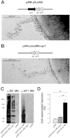
Adhesive properties of L. lactis expressing the FCT-1 M6 pilus operon and its Δap1_M6 derivative. A. Immunoelectron microscopy image of a L. lactis recombinant strain harbouring the entire FCT-1 M6 pilus operon. B. Immunoelectron microscopy image of a L. lactis recombinant strain harbouring the FCT-1 M6 pilus operon deprived of the ap1 gene. In both cases, bacteria were first incubated with polyclonal sera raised against rBP_M6, and then labelled with secondary antibodies conjugated with 5 nm gold particles. Pictures were taken at 60× magnification. C. Immunoblot analysis of L. lactis cell wall extracts incubated with antisera raised against rBP_M6 and rAP1_M6 pilus proteins. D. Binding of L. lactis recombinant strains to A549 lung epithelial cells (moi 20:1) after 30 min of incubation, expressed as percentage of cell-associated versus total bacteria. Results are presented as the mean and standard deviation values of three independent experiments run in triplicate wells. Significant differences between strains (**P < 0.01) were estimated using the Student's t-test.
Fig 3
Fig 3
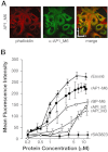
Binding of rAP1_M6 and other recombinant proteins to epithelial cells. A. Confocal microscopy analysis of rAP1_M6 protein stained with specific antibodies (green) binding to epithelial cells (labelled with phalloidin, red); the x- and y-axis arrows in the merged image indicate 27 µm dimensions. B. FACS analysis of GAS proteins binding to A549 cells. Cells were incubated with increasing concentrations of the following recombinant proteins: pilus ancillary proteins rAP1_M6, rAP1_M1 and rAP1_M3, the pilus backbone rBP_M6, the rEmm6 protein and, as negative control, rSAG0823 from S. agalactiae. Cell-bound proteins were detected with specific antibodies and fluorescent secondary antisera, followed by flow cytometry analysis. Results are presented as the mean and standard deviation values of three independent experiments.
Fig 6
Fig 6
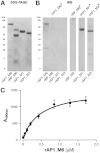
Biochemical analysis of protein–protein interactions involving pilus proteins. A. SDS-PAGE analysis of recombinant pilus proteins rAP1_M6, rBP_M6, rAP1_M1 and rAP1_M3; lane 2 contains a main band of 62 kDa corresponding to rBP_M6 and a secondary band of about 90 kDa, which corresponds to an impurity from E. coli. B. Far Western blot analysis using biotinylated rAP1_M6 (rAP1_M6*) as probe against the recombinant proteins shown in (A); Far Western blot assay of biotinylated rBP_M6*, rAP1_M1* and rAP1_M3* as probes against the respective pilus proteins loaded on SDS-PAGE. C. ELISA saturation kinetic analysis of biotinylated rAP1_M6* protein bound to immobilized unlabelled rAP1_M6. All experiments were conducted in triplicate with reproducible results, and mean and standard deviation values are reported.
Fig 4
Fig 4
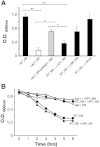
Role of AP1_M6 in biofilm formation and bacterial aggregation. A. Crystal violet biofilm formation assay. GAS WT_M6, Δap1_M6 and complemented bacteria were grown for 10 h in 24-well plates, washed with PBS and stained with crystal violet. Experiments with WT_M6 were also conducted in the presence of purified mouse polyclonal antibodies raised against rAP1_M6 and rBP_M6 pilus components, and against rScpA as control. The mean and SD values of three independent experiments run in triplicate wells are reported. Significant differences between strains calculated by Student's t-test are shown by asterisks, **P < 0.01, *P < 0.05. B. Sedimentation of GAS WT_M6 grown in the presence or absence of 1 µM rAP1_M6 or rBP_M6, compared with that of Δap1_M6 mutant strain. Bacteria were grown for 16 h, bacterial precipitates were suspended by mixing, and OD values were at 600 nm measured at different time intervals under static conditions; the presented data derive from one of three experiments with reproducible results.
Fig 5
Fig 5
Flow-cytometry analysis of the interaction between recombinant rAP1_M6 and GAS or Lactococcus bacteria. A. FACS analysis of GAS WT_M6, its deletion mutant Δap1_M6 and the complemented strain Δap1_M6(pAM ap1_M6). B. FACS analysis of L. lactis carrying either an empty vector (pAM), or a plasmid containing the FCT-1 M6 pilus operon (pAM-pilus M6) or the same operon except for a deletion in the ap1_M6 gene. In all cases bacteria were incubated with 5 µg of biotinylated rAP1_M6 and binding of the protein to the cells was revealed by R-Phycoerythrin-conjugated Streptavidin. Black histograms indicate staining of bacteria with streptavidin alone, while red histograms correspond to bacteria incubated with rAP1_M6 protein and then streptavidin. Binding was calculated by subtracting the mean fluorescence of bacteria stained with biotinylated rAP1_M6 protein from that of the unstained bacteria (ΔMFI). The average ΔMFI value from three independent experiments is shown.
Fig 7
Fig 7
Analysis of bacterial infection in mice and blood survival of WT_M6 and Δap1_M6 strains. A. Number of bacteria obtained after plating dissected and homogenized spleens, lungs, kidneys (total number of cfu) and blood (cfu ml−1) collected 24 h post intraperitoneal infection of mice using 2 × 10⁷ cfu of GAS WT_M6 or Δap1_M6 strains (12 animals per strain, in three independent experiments with four animals each). Each circle represents the number of bacteria recovered in the organ of one mouse, and the average number of bacteria from all mice is indicated by a bar. Differences were statistically significant in all organs (**P < 0.01, U-test). B. Competitive index of GAS WT_M6 versus Δap1_M6 strains. Eight mice (in two independent experiments with four animals each) were infected intraperitoneally with 107 cfu of WT_M6 and 107 cfu of Δap1_M6. After 24 h, bacteria were recovered from the lungs and spleens of sacrificed animals and plated. Wild-type bacteria were discriminated from mutant strains by colony blot analysis using antibodies to rAP1_M6 protein. Each circle represents the ratio between the number of cfu of the two strains in the organ of one mouse, and the average ratio is indicated by a bar. C. Survival of GAS WT_M6 with or without rAP1_M6, of the deletion mutant Δap1_M6, and of the complemented strain Δap1_M6(pAMap1_M6) in whole blood from rabbits. For each strain, the average relative growth rate (cfu at time 5 h divided by cfu at time 0) versus that of wild type (to which we assigned a value of 100) obtained from three different experiments, and its standard deviation, are reported. Significant differences between strains were calculated by Student's t-test, **P < 0.01, *P < 0.05.

Similar articles

Cited by

References

    1. Abbot EL, Smith WD, Siou G, Chiriboga C, Smith RJ, Wilson JA, et al. Pili mediate specific adhesion of Streptococcus pyogenes to human tonsil and skin. Cell Microbiol. 2007;9:1822–1833. - PubMed
    1. Banerjee A, Kim BJ, Carmona EM, Cutting AS, Gurney MA, Carlos C, et al. Bacterial pili exploit integrin machinery to promote immune activation and efficient blood-brain barrier penetration. Nat Commun. 2011;2:462. - PMC - PubMed
    1. Barocchi MA, Ries J, Zogaj X, Hemsley C, Albiger B, Kanth A, et al. A pneumococcal pilus influences virulence and host inflammatory responses. Proc Natl Acad Sci USA. 2006;103:2857–2862. - PMC - PubMed
    1. Bessen DE, Kalia A. Genomic localization of a T serotype locus to a recombinatorial zone encoding extracellular matrix-binding proteins in Streptococcus pyogenes. Infect Immun. 2002;70:1159–1167. - PMC - PubMed
    1. Buccato S, Maione D, Rinaudo CD, Volpini G, Taddei AR, Rosini R, et al. Use of Lactococcus lactis expressing pili from group B Streptococcus as a broad-coverage vaccine against streptococcal disease. J Infect Dis. 2006;194:331–340. - PubMed